Division of Medical Oncology, Department of Internal Medicine, St. Vincent's Hospital, College of Medicine, The Catholic University of Korea, Seoul, Korea (the Republic of).
Department of Surgery, Yonsei University College of Medicine, Seoul, Korea (the Republic of).
J Immunother Cancer. 2021 Oct;9(10). doi: 10.1136/jitc-2021-003353.
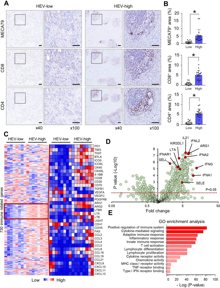
High endothelial venule (HEV) is a specialized vasculature for lymphocyte trafficking. While HEVs are frequently observed within gastric cancer (GC), the vascular-immune interaction between HEV and tumor-infiltrating lymphocytes (TILs) has not been well elucidated. In this study, we aimed to unveil the potential value of HEVs as a surrogate marker for T-cell inflamed immune microenvironment in GC using a large number of prospectively collected surgical specimens of GC.
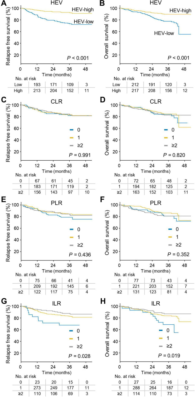
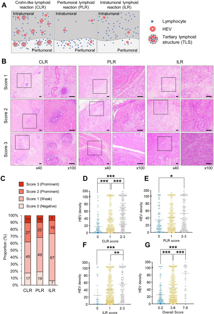
We included 460 patients with GC who underwent surgical resection. Nanostring PanCancer immune profiling was performed to evaluate the immunological phenotype of GCs. HEV density and three distinct patterns of TILs (Crohn-like lymphoid reaction, peritumoral lymphoid reaction, and intratumoral lymphoid reaction) were analyzed for their relationship and evaluated as prognostic factors for relapse-free survival (RFS) and overall survival (OS).
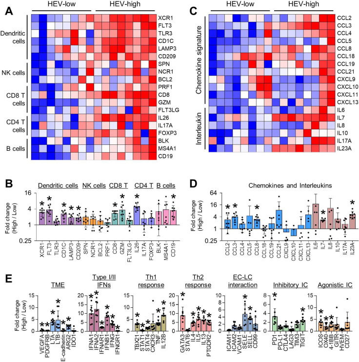
HEV-high GC revealed increased infiltration by immune cell subsets, including dendritic cells, CD8 cytotoxic T cells, and CD4 helper T cells. In addition, HEV-high GC demonstrated increased immune-modulating chemokines, type I or II interferon pathway, and immune checkpoints, all of which indicate the inflamed tumor microenvironment (TME). All three distinct patterns of TILs were associated with HEV density. In survival analysis, patients with HEV-high GC displayed significantly longer RFS and OS than those with HEV-low GC (p<0.001 for RFS, p<0.001 for OS). Multivariate analysis demonstrated that HEV was the most significant immunological prognostic factor for RFS (patients with high HEV compared with those with low HEV; HR 0.412, 95% CI 0.241 to 0.705, p=0.001) and OS (HR 0.547, 95% CI 0.329 to 0.909, p=0.02) after adjustment for age, stage, and TIL.
HEV is the most significant immunological prognosticator for RFS and OS in resected GC, indicating inflamed TME.
高内皮小静脉(HEV)是淋巴细胞运输的特化血管。虽然 HEV 常在胃癌(GC)中观察到,但 HEV 与肿瘤浸润淋巴细胞(TIL)之间的血管免疫相互作用尚未得到充分阐明。在这项研究中,我们旨在使用大量前瞻性收集的 GC 手术标本,揭示 HEV 作为 GC 中 T 细胞炎症免疫微环境替代标志物的潜在价值。
我们纳入了 460 名接受手术切除的 GC 患者。采用 Nanostring PanCancer 免疫分析技术评估 GC 的免疫表型。分析 HEV 密度和三种不同类型的 TIL(克罗恩样淋巴反应、肿瘤周围淋巴反应和肿瘤内淋巴反应),以研究它们之间的关系,并将其评估为无复发生存(RFS)和总生存(OS)的预后因素。
HEV 高 GC 显示免疫细胞亚群浸润增加,包括树突状细胞、CD8 细胞毒性 T 细胞和 CD4 辅助 T 细胞。此外,HEV 高 GC 表现出增加的免疫调节趋化因子、I 型或 II 型干扰素途径和免疫检查点,所有这些都表明存在炎症肿瘤微环境(TME)。三种不同类型的 TIL 均与 HEV 密度相关。在生存分析中,HEV 高 GC 患者的 RFS 和 OS 明显长于 HEV 低 GC 患者(RFS 方面,p<0.001;OS 方面,p<0.001)。多变量分析表明,HEV 是 RFS(与低 HEV 相比,高 HEV 患者;HR 0.412,95%CI 0.241 至 0.705,p=0.001)和 OS(HR 0.547,95%CI 0.329 至 0.909,p=0.02)最重要的免疫预后因素,调整年龄、分期和 TIL 后。
HEV 是 GC 患者 RFS 和 OS 的最重要的免疫预后指标,表明存在炎症 TME。