Queensland University of Technology, School of Biomedical Sciences, Faculty of Health, Brisbane, Australia
The University of Queensland Diamantina Institute, The University of Queensland, Woollongabba, Australia.
Eur Respir J. 2022 Jun 2;59(6). doi: 10.1183/13993003.01881-2021. Print 2022 Jun.
The severe acute respiratory syndrome coronavirus 2 (SARS-CoV-2) which emerged in late 2019 has spread globally, causing a pandemic of respiratory illness designated coronavirus disease 2019 (COVID-19). A better definition of the pulmonary host response to SARS-CoV-2 infection is required to understand viral pathogenesis and to validate putative COVID-19 biomarkers that have been proposed in clinical studies.
Here, we use targeted transcriptomics of formalin-fixed paraffin-embedded tissue using the NanoString GeoMX platform to generate an in-depth picture of the pulmonary transcriptional landscape of COVID-19, pandemic H1N1 influenza and uninfected control patients.
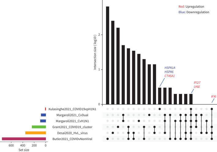
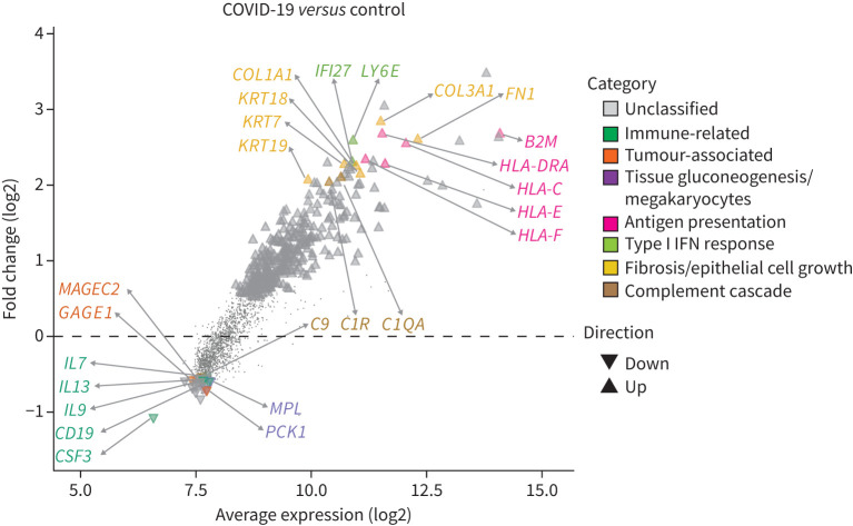
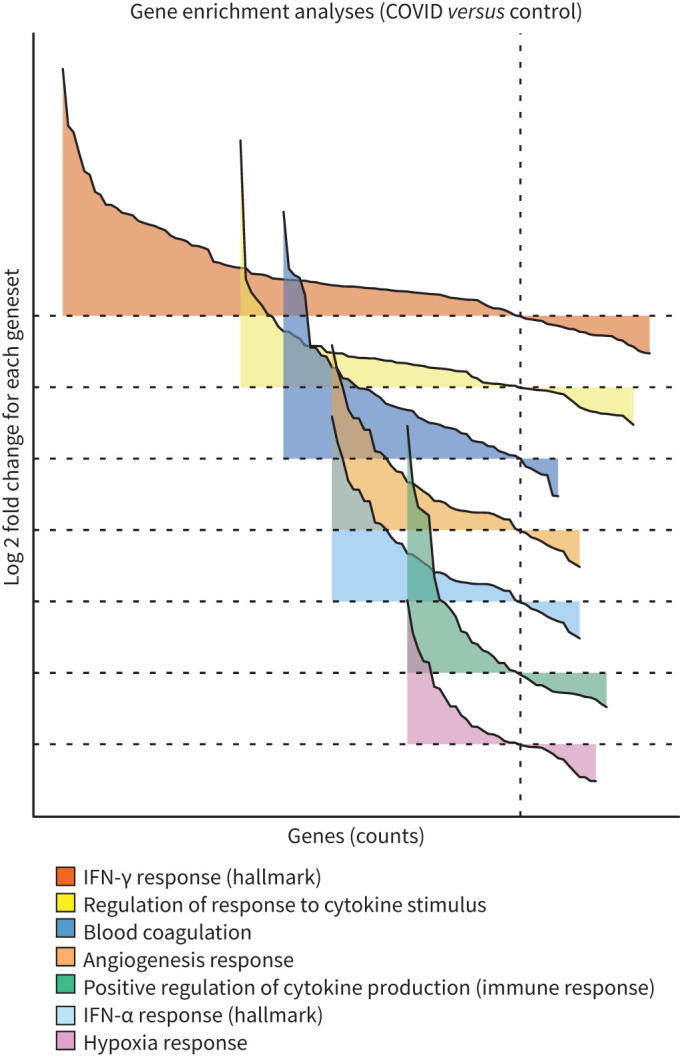
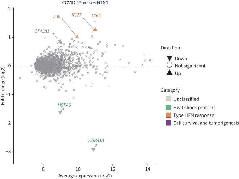
Host transcriptomics showed a significant upregulation of genes associated with inflammation, type I interferon production, coagulation and angiogenesis in the lungs of COVID-19 patients compared to non-infected controls. SARS-CoV-2 was non-uniformly distributed in lungs (emphasising the advantages of spatial transcriptomics) with the areas of high viral load associated with an increased type I interferon response. Once the dominant cell type present in the sample, within patient correlations and patient-patient variation, had been controlled for, only a very limited number of genes were differentially expressed between the lungs of fatal influenza and COVID-19 patients. Strikingly, the interferon-associated gene , previously identified as a useful blood biomarker to differentiate bacterial and viral lung infections, was significantly upregulated in the lungs of COVID-19 patients compared to patients with influenza.
Collectively, these data demonstrate that spatial transcriptomics is a powerful tool to identify novel gene signatures within tissues, offering new insights into the pathogenesis of SARS-COV-2 to aid in patient triage and treatment.
2019 年末出现的严重急性呼吸综合征冠状病毒 2(SARS-CoV-2)已在全球范围内传播,导致被指定为 2019 年冠状病毒病(COVID-19)的呼吸道疾病大流行。为了更好地了解病毒发病机制并验证临床研究中提出的假定 COVID-19 生物标志物,需要更深入地了解 SARS-CoV-2 感染对肺部宿主的反应。
在这里,我们使用 NanoString GeoMX 平台对福尔马林固定石蜡包埋组织进行靶向转录组学分析,以生成 COVID-19、大流行性 H1N1 流感和未感染对照患者肺部转录组学全景图。
宿主转录组学显示,与未感染对照相比,COVID-19 患者肺部与炎症、I 型干扰素产生、凝血和血管生成相关的基因显著上调。SARS-CoV-2 在肺部的分布不均匀(强调空间转录组学的优势),高病毒载量区域与 I 型干扰素反应增加有关。在控制了样本中存在的主要细胞类型、患者内相关性和患者间变异后,只有少数基因在致命性流感和 COVID-19 患者的肺部之间存在差异表达。引人注目地是,干扰素相关基因,先前被鉴定为区分细菌和病毒肺部感染的有用血液生物标志物,在 COVID-19 患者的肺部中显著上调,与流感患者相比。
总的来说,这些数据表明,空间转录组学是一种识别组织内新基因特征的强大工具,为 SARS-COV-2 的发病机制提供了新的见解,有助于患者分诊和治疗。