• 文献检索
  • 文档翻译
  • 深度研究
  • 学术资讯
  • Suppr Zotero 插件Zotero 插件
  • 邀请有礼
  • 套餐&价格
  • 历史记录
应用&插件
Suppr Zotero 插件Zotero 插件浏览器插件Mac 客户端Windows 客户端微信小程序
定价
高级版会员购买积分包购买API积分包
服务
文献检索文档翻译深度研究API 文档MCP 服务
关于我们
关于 Suppr公司介绍联系我们用户协议隐私条款
关注我们

Suppr 超能文献

核心技术专利:CN118964589B侵权必究
粤ICP备2023148730 号-1Suppr @ 2026

文献检索

告别复杂PubMed语法,用中文像聊天一样搜索,搜遍4000万医学文献。AI智能推荐,让科研检索更轻松。

立即免费搜索

文件翻译

保留排版,准确专业,支持PDF/Word/PPT等文件格式,支持 12+语言互译。

免费翻译文档

深度研究

AI帮你快速写综述,25分钟生成高质量综述,智能提取关键信息,辅助科研写作。

立即免费体验

一种用于光镜下鉴定犬肥大细胞的双重组织化学/免疫组织化学染色法。

A Double Histochemical/Immunohistochemical Staining for the Identification of Canine Mast Cells in Light Microscopy.

作者信息

Gobbo Francesca, Sarli Giuseppe, De Silva Margherita, Galiazzo Giorgia, Chiocchetti Roberto, Morini Maria

机构信息

Department of Veterinary Medical Sciences, University of Bologna, Ozzano dell'Emilia, 40064 Bologna, Italy.

出版信息

Vet Sci. 2021 Oct 12;8(10):229. doi: 10.3390/vetsci8100229.

DOI:10.3390/vetsci8100229
PMID:34679059
原文链接:https://pmc.ncbi.nlm.nih.gov/articles/PMC8537840/
Abstract

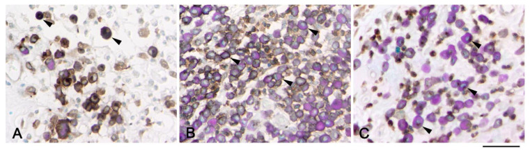
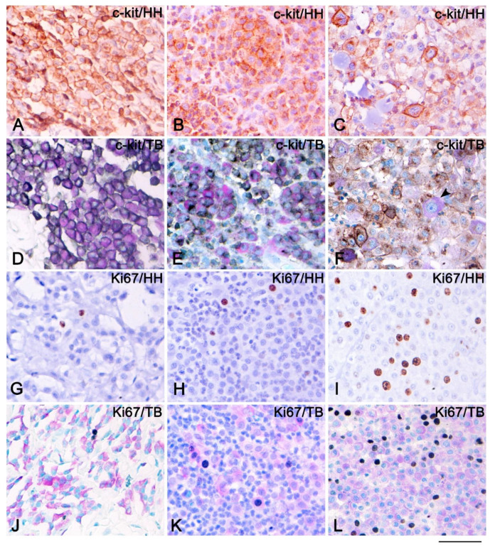
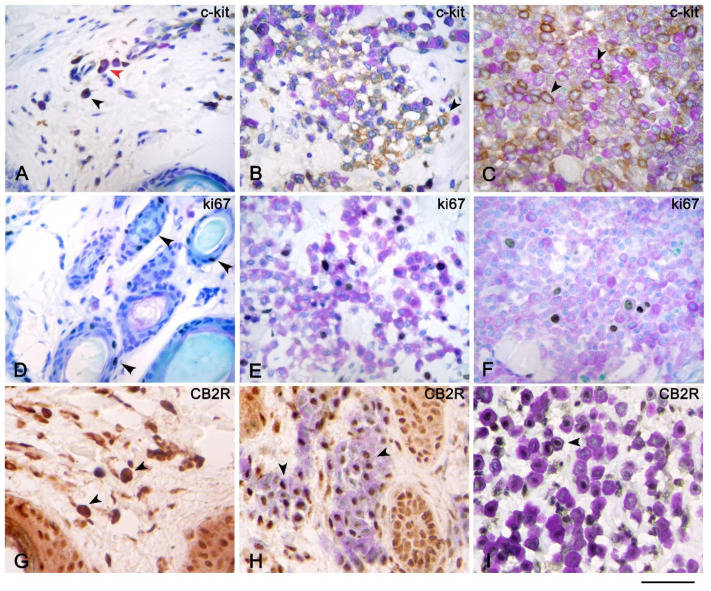
Immunohistochemistry (IHC) is a widely used technique in diagnostic pathology, but the simultaneous analysis of more than one antibody at a time with different chromogens is rather complex, time-consuming, and quite expensive. In order to facilitate the identification of mast cells (MCs) during immunohistochemical analysis of membrane and/or nuclear markers, we propose a new staining method that includes the association of IHC and toluidine blue as a counterstain. To achieve this goal, we tested c-kit, Ki67, and cannabinoid receptor 2 on several cases of cutaneous canine mast cell tumors (MCTs), cutaneous mastocytosis, and atopic dermatitis. The results obtained show how this double staining technique, although limited to non-cytoplasmic markers and of little use in poorly differentiated MCTs in which MC metachromasia is hard to see, can be used during the evaluation of nuclear and/or membranous immunohistochemical markers in all canine cutaneous disorders, especially if characterized by the presence of a low number of MCs. It can help to evaluate those MCTs in which neoplastic MCs must be clearly distinguished from inflammatory cells that can infiltrate the tumor itself, in facilitating the calculation of the Ki67 index. Moreover, it can be used to study the expression of new markers in both animal and human tissues containing MCs and in MC disorders.

摘要

免疫组织化学(IHC)是诊断病理学中广泛使用的技术,但同时使用不同显色剂对多种抗体进行分析相当复杂、耗时且成本高昂。为了在对膜和/或核标记物进行免疫组织化学分析时便于识别肥大细胞(MCs),我们提出了一种新的染色方法,该方法包括将免疫组织化学与甲苯胺蓝作为复染剂相结合。为实现这一目标,我们在几例犬皮肤肥大细胞瘤(MCTs)、皮肤肥大细胞增多症和特应性皮炎病例中检测了c-kit、Ki67和大麻素受体2。获得的结果表明,这种双重染色技术虽然仅限于非细胞质标记物,且在难以观察到MC异染性的低分化MCTs中作用不大,但可用于评估所有犬皮肤疾病中的核和/或膜免疫组织化学标记物,特别是在MC数量较少的情况下。它有助于评估那些必须将肿瘤性MC与可浸润肿瘤本身的炎性细胞清楚区分的MCTs,有助于计算Ki67指数。此外,它可用于研究含有MCs的动物和人类组织以及MC疾病中新标记物的表达。

https://cdn.ncbi.nlm.nih.gov/pmc/blobs/e425/8537840/0f6f646b7645/vetsci-08-00229-g003.jpg
https://cdn.ncbi.nlm.nih.gov/pmc/blobs/e425/8537840/148526fcb4aa/vetsci-08-00229-g001.jpg
https://cdn.ncbi.nlm.nih.gov/pmc/blobs/e425/8537840/f19dd39f8dcf/vetsci-08-00229-g002.jpg
https://cdn.ncbi.nlm.nih.gov/pmc/blobs/e425/8537840/0f6f646b7645/vetsci-08-00229-g003.jpg
https://cdn.ncbi.nlm.nih.gov/pmc/blobs/e425/8537840/148526fcb4aa/vetsci-08-00229-g001.jpg
https://cdn.ncbi.nlm.nih.gov/pmc/blobs/e425/8537840/f19dd39f8dcf/vetsci-08-00229-g002.jpg
https://cdn.ncbi.nlm.nih.gov/pmc/blobs/e425/8537840/0f6f646b7645/vetsci-08-00229-g003.jpg

相似文献

1
A Double Histochemical/Immunohistochemical Staining for the Identification of Canine Mast Cells in Light Microscopy.一种用于光镜下鉴定犬肥大细胞的双重组织化学/免疫组织化学染色法。
Vet Sci. 2021 Oct 12;8(10):229. doi: 10.3390/vetsci8100229.
2
Cellular proliferation in canine cutaneous mast cell tumors: associations with c-KIT and its role in prognostication.犬皮肤肥大细胞瘤中的细胞增殖:与c-KIT的关联及其在预后评估中的作用
Vet Pathol. 2007 May;44(3):298-308. doi: 10.1354/vp.44-3-298.
3
Ki67/KIT double immunohistochemical staining in cutaneous mast cell tumors from Boxer dogs.犬皮肤肥大细胞瘤的 Ki67/KIT 双重免疫组织化学染色。
Res Vet Sci. 2015 Oct;102:122-6. doi: 10.1016/j.rvsc.2015.08.007. Epub 2015 Aug 8.
4
The use of KIT and tryptase expression patterns as prognostic tools for canine cutaneous mast cell tumors.将KIT和类胰蛋白酶表达模式用作犬皮肤肥大细胞瘤预后工具的研究
Vet Pathol. 2004 Jul;41(4):371-7. doi: 10.1354/vp.41-4-371.
5
Immunohistochemical application of an antibody specific for human CD1a to the diagnosis of canine mast cell tumour.一种针对人CD1a的特异性抗体在犬肥大细胞瘤诊断中的免疫组织化学应用。
J Comp Pathol. 2008 Jul;139(1):40-6. doi: 10.1016/j.jcpa.2008.05.001. Epub 2008 Jul 7.
6
Expression of KIT receptor in feline cutaneous mast cell tumors.KIT受体在猫皮肤肥大细胞瘤中的表达。
Vet Pathol. 2009 Sep;46(5):878-83. doi: 10.1354/vp.08-VP-0084-R-FL. Epub 2009 May 9.
7
Immunohistochemical and Molecular Genetic Analysis of Canine Digital Mast Cell Tumours.犬趾部肥大细胞肿瘤的免疫组织化学和分子遗传学分析
Animals (Basel). 2023 May 19;13(10):1694. doi: 10.3390/ani13101694.
8
HSP32 and HSP90 Immunoexpression, in Relation to Kit Pattern, Grading, and Mitotic Count in Canine Cutaneous Mast Cell Tumors.犬皮肤肥大细胞瘤中HSP32和HSP90免疫表达与Kit模式、分级及有丝分裂计数的关系
Vet Pathol. 2017 Mar;54(2):222-225. doi: 10.1177/0300985816669405. Epub 2016 Sep 29.
9
c-KIT messenger RNA and protein expression and mutations in canine cutaneous mast cell tumors: correlations with post-surgical prognosis.犬皮肤肥大细胞瘤中c-KIT信使核糖核酸和蛋白质表达及突变:与术后预后的相关性
J Vet Diagn Invest. 2012 Jan;24(1):116-26. doi: 10.1177/1040638711425945. Epub 2011 Dec 6.
10
Evaluation of the kinase domain of c-KIT in canine cutaneous mast cell tumors.犬皮肤肥大细胞瘤中c-KIT激酶结构域的评估
BMC Cancer. 2006 Apr 1;6:85. doi: 10.1186/1471-2407-6-85.

引用本文的文献

1
Oral treatment with fructus extract modulates mast cells in canine atopic dermatitis.用果实提取物进行口服治疗可调节犬特应性皮炎中的肥大细胞。
Front Vet Sci. 2025 Apr 9;12:1531313. doi: 10.3389/fvets.2025.1531313. eCollection 2025.
2
-Lyophilized Fruit Extract Attenuated Acute Redox-Homeostatic Imbalance via Mutagenic and Oxidative Stress Modulation in Mice Model on Gentamicin-Induced Nephrotoxicity.冻干水果提取物通过调节庆大霉素诱导的肾毒性小鼠模型中的诱变和氧化应激,减轻急性氧化还原稳态失衡。
Pharmaceuticals (Basel). 2025 Jan 13;18(1):85. doi: 10.3390/ph18010085.
3
Cannabinoid receptors in the inflammatory cells of canine atopic dermatitis.

本文引用的文献

1
Histological classification and expression of markers of canine mast cell tumors.犬肥大细胞瘤的组织学分类及标志物表达
Vet World. 2020 Aug;13(8):1627-1634. doi: 10.14202/vetworld.2020.1627-1634. Epub 2020 Aug 18.
2
Overview of multiplex immunohistochemistry/immunofluorescence techniques in the era of cancer immunotherapy.癌症免疫治疗时代的多重免疫组化/免疫荧光技术概述。
Cancer Commun (Lond). 2020 Apr;40(4):135-153. doi: 10.1002/cac2.12023. Epub 2020 Apr 17.
3
Diagnostic immunohistochemistry for canine cutaneous round cell tumours - retrospective analysis of 60 cases.
犬特应性皮炎炎症细胞中的大麻素受体
Front Vet Sci. 2022 Sep 15;9:987132. doi: 10.3389/fvets.2022.987132. eCollection 2022.
犬皮肤圆形细胞瘤的诊断性免疫组化——60例回顾性分析
Folia Histochem Cytobiol. 2019;57(3):146-154. doi: 10.5603/FHC.a2019.0016. Epub 2019 Sep 25.
4
Mast cell chymase: morphofunctional characteristics.肥大细胞糜酶:形态功能特征。
Histochem Cell Biol. 2019 Oct;152(4):253-269. doi: 10.1007/s00418-019-01803-6. Epub 2019 Aug 8.
5
Prognostic and predictive significance of KIT protein expression and c-kit gene mutation in canine cutaneous mast cell tumours: A consensus of the Oncology-Pathology Working Group.犬皮肤肥大细胞瘤中 KIT 蛋白表达和 c-kit 基因突变的预后和预测意义:肿瘤-病理学工作组的共识。
Vet Comp Oncol. 2019 Dec;17(4):451-455. doi: 10.1111/vco.12518. Epub 2019 Jul 9.
6
Diagnosis and Prognosis of Canine Cutaneous Mast Cell Tumors.犬皮肤肥大细胞瘤的诊断与预后
Vet Clin North Am Small Anim Pract. 2019 Sep;49(5):819-836. doi: 10.1016/j.cvsm.2019.04.002. Epub 2019 Jun 6.
7
Update on pathogenesis, diagnosis, and treatment of atopic dermatitis in dogs.犬特应性皮炎的发病机制、诊断与治疗进展
J Am Vet Med Assoc. 2019 Jun 1;254(11):1291-1300. doi: 10.2460/javma.254.11.1291.
8
The Endocannabinoid System as a Target in Cancer Diseases: Are We There Yet?内源性大麻素系统作为癌症疾病的一个靶点:我们做到了吗?
Front Pharmacol. 2019 Apr 5;10:339. doi: 10.3389/fphar.2019.00339. eCollection 2019.
9
Immunofluorescence staining protocol for STED nanoscopy of Plasmodium-infected red blood cells.疟原虫感染红细胞的受激发射损耗纳米显微镜免疫荧光染色方案。
Mol Biochem Parasitol. 2019 Apr;229:47-52. doi: 10.1016/j.molbiopara.2019.02.007. Epub 2019 Mar 1.
10
An Introduction to Performing Immunofluorescence Staining.免疫荧光染色操作介绍。
Methods Mol Biol. 2019;1897:299-311. doi: 10.1007/978-1-4939-8935-5_26.