Kim Min Jeong, Kang Hyun-Hee, Seo Yeung Jin, Kim Kyung-Min, Kim Young-Jun, Jung Sung Keun
School of Food Science and Biotechnology, Kyungpook National University, Daegu 41566, Korea.
Department of Food Science and Technology, Seoul National University of Science and Technology, Seoul 01811, Korea.
Antioxidants (Basel). 2021 Sep 23;10(10):1507. doi: 10.3390/antiox10101507.
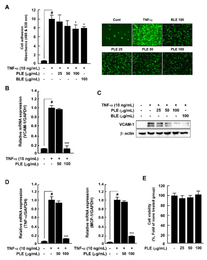
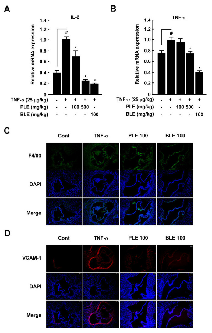
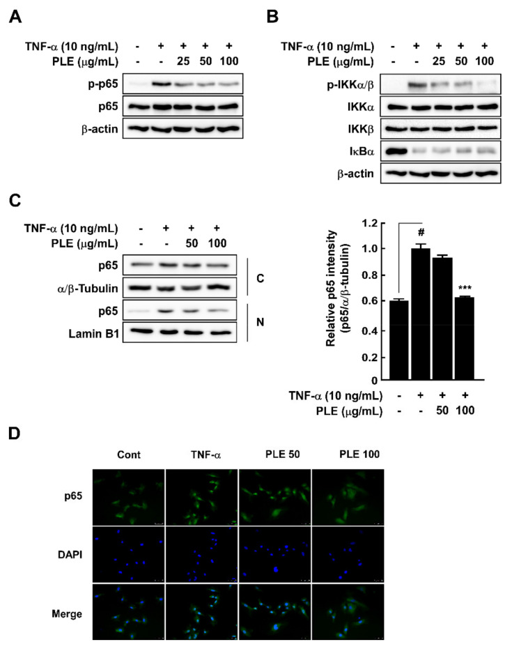
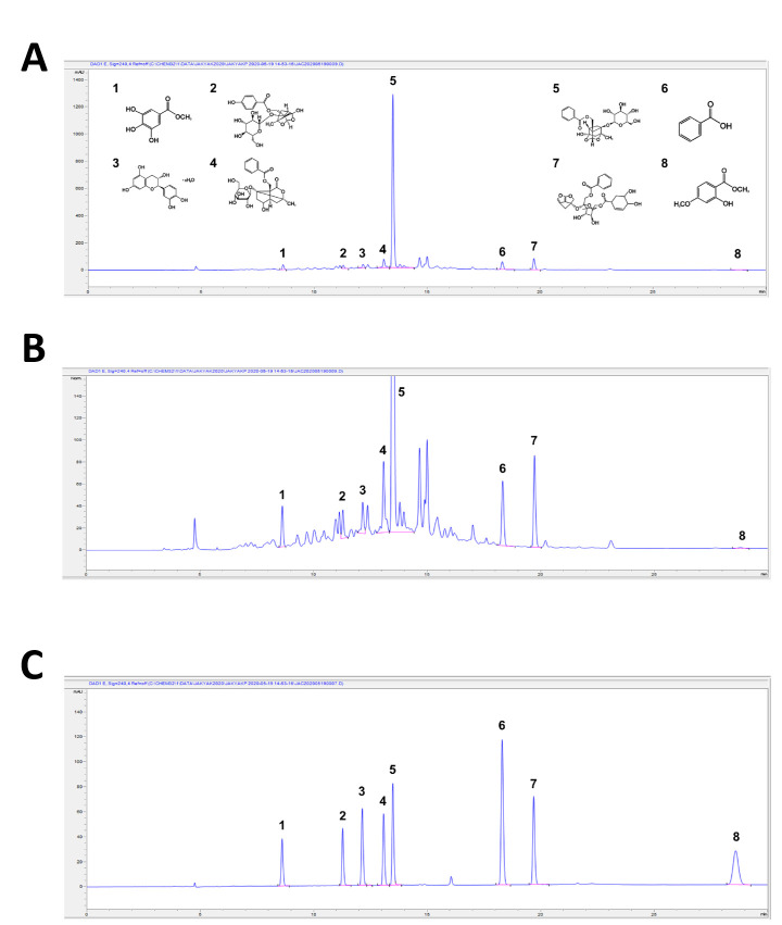
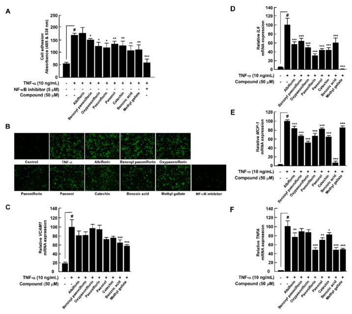
Although various physiological activities of compounds obtained from have been reported, the effects of extract (PLE) on early atherosclerosis remain unclear. Therefore, in this study, we investigated the in vitro and in vivo antiatherosclerosis and in vitro antioxidant effects of PLE and its compounds. PLE suppresses the tumor necrosis factor (TNF)-α-induced capacity of THP-1 cells to adhere to human umbilical vein endothelial cells (HUVECs), vascular cell adhesion molecule (VCAM)-1 expression, and nuclear factor kappa-light-chain-enhancer of activated B cells (NF-κB) signaling in HUVECs. PLE also suppresses TNF-α-induced nuclear translocation of NF-κB p65 from cytosol as well as the enhanced and C-C motif chemokine ligand 2 (CCL2) mRNA expression in HUVECs. We identified and quantified the following PLE compounds using high-performance liquid chromatography with diode array detection: methyl gallate, oxypaeoniflorin, catechin, albiflorin, paeoniflorin, benzoic acid, benzoylpaeoniflorin, and paeonol. Among these, methyl gallate had the strongest inhibitory effect on monocyte adherence to TNF-α-induced HUVECs and the VCAM-1 expression. Reverse transcriptase real-time quantitative polymerase chain reaction showed that PLE compounds had a dissimilar inhibition effect on TNF-α-induced mRNA expression levels of , , and in HUVECs. Except for paeonol, the compounds inhibited lipopolysaccharide (LPS)-induced reactive oxygen species production in RAW264.7 cells. In vivo, oral administration of PLE improved TNF-α-induced macrophage infiltration to the vascular endothelium and expression of VCAM-1, as well as and gene expression in the main artery of mice. PLE could be useful as a nutraceutical material against early atherosclerosis via the combined effects of its components.
尽管已报道了从[具体来源未提及]获得的化合物的各种生理活性,但[具体植物名称]提取物(PLE)对早期动脉粥样硬化的影响仍不清楚。因此,在本研究中,我们研究了PLE及其化合物的体外和体内抗动脉粥样硬化作用以及体外抗氧化作用。PLE可抑制肿瘤坏死因子(TNF)-α诱导的THP-1细胞与人脐静脉内皮细胞(HUVECs)黏附的能力、血管细胞黏附分子(VCAM)-1的表达以及HUVECs中活化B细胞核因子κB(NF-κB)信号通路。PLE还可抑制TNF-α诱导的NF-κB p65从细胞质向细胞核的转位以及HUVECs中[具体基因未提及]和C-C基序趋化因子配体2(CCL2)mRNA表达的增强。我们使用带二极管阵列检测的高效液相色谱法鉴定并定量了以下PLE化合物:没食子酸甲酯、氧化芍药苷、儿茶素、白花芍药苷、芍药苷、苯甲酸、苯甲酰芍药苷和丹皮酚。其中,没食子酸甲酯对单核细胞黏附于TNF-α诱导的HUVECs和VCAM-1表达的抑制作用最强。逆转录酶实时定量聚合酶链反应表明,PLE化合物对TNF-α诱导的HUVECs中[具体基因未提及]、[具体基因未提及]和[具体基因未提及]的mRNA表达水平具有不同的抑制作用。除丹皮酚外,这些化合物均抑制脂多糖(LPS)诱导的RAW264.7细胞中活性氧的产生。在体内,口服PLE可改善TNF-α诱导的巨噬细胞向血管内皮的浸润以及VCAM-1的表达,以及小鼠主动脉中[具体基因未提及]和[具体基因未提及]基因的表达。通过其成分的联合作用,PLE可能作为一种抗早期动脉粥样硬化的营养保健品。