Chong Gun Oh, Park Shin-Hyung, Park Nora Jee-Young, Bae Bong Kyung, Lee Yoon Hee, Jeong Shin Young, Kim Jae-Chul, Park Ji Young, Ando Yu, Han Hyung Soo
Department of Obstetrics and Gynecology, School of Medicine, Kyungpook National University, Daegu 41944, Korea.
Department of Obstetrics and Gynecology, Kyungpook National University Chilgok Hospital, Daegu 41944, Korea.
Cancers (Basel). 2021 Oct 14;13(20):5140. doi: 10.3390/cancers13205140.
Our previous study demonstrated that tumor budding (TB) status was associated with inferior overall survival in cervical cancer. The purpose of this study is to evaluate whether radiomic features can predict TB status in cervical cancer patients.
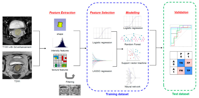
Seventy-four patients with cervical cancer who underwent preoperative MRI and radical hysterectomy from 2011 to 2015 at our institution were enrolled. The patients were randomly allocated to the training dataset ( = 48) and test dataset ( = 26). Tumors were segmented on axial gadolinium-enhanced T1- and T2-weighted images. A total of 2074 radiomic features were extracted. Four machine learning classifiers, including logistic regression (LR), random forest (RF), support vector machine (SVM), and neural network (NN), were used. The trained models were validated on the test dataset.
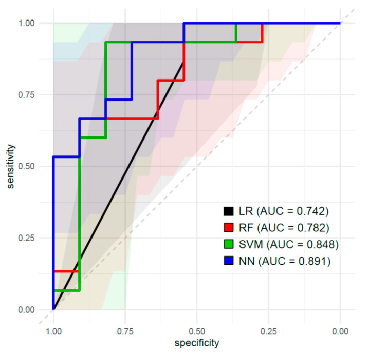
Twenty radiomic features were selected; all were features from filtered-images and 85% were texture-related features. The area under the curve values and accuracy of the models by LR, RF, SVM and NN were 0.742 and 0.769, 0.782 and 0.731, 0.849 and 0.885, and 0.891 and 0.731, respectively, in the test dataset.
MRI-based radiomic features could predict TB status in patients with cervical cancer.
我们之前的研究表明,肿瘤芽生(TB)状态与宫颈癌患者较差的总生存期相关。本研究的目的是评估影像组学特征是否能够预测宫颈癌患者的TB状态。
纳入了2011年至2015年在我院接受术前MRI检查和根治性子宫切除术的74例宫颈癌患者。患者被随机分配到训练数据集(n = 48)和测试数据集(n = 26)。在轴位钆增强T1加权和T2加权图像上对肿瘤进行分割。共提取了2074个影像组学特征。使用了四种机器学习分类器,包括逻辑回归(LR)、随机森林(RF)、支持向量机(SVM)和神经网络(NN)。在测试数据集上对训练好的模型进行验证。
选择了20个影像组学特征;所有特征均来自滤波后的图像,85%为纹理相关特征。在测试数据集中,LR、RF、SVM和NN模型的曲线下面积值和准确率分别为0.742和0.769、0.782和0.731、0.849和0.885、0.891和0.731。
基于MRI的影像组学特征能够预测宫颈癌患者的TB状态。