Gültekin Emin, Wetz Christoph, Braun Jürgen, Geisel Dominik, Furth Christian, Hamm Bernd, Sack Ingolf, Marticorena Garcia Stephan R
Department of Radiology, Campus Virchow Klinikum, Charité-Universitätsmedizin Berlin, Corporate Member of Freie Universität Berlin, Humboldt-Universität zu Berlin, and Berlin Institute of Health, Charitéplatz 1, 10117 Berlin, Germany.
Department of Nuclear Medicine, Charité-Universitätsmedizin Berlin, Corporate Member of Freie Universität Berlin, Humboldt-Universität zu Berlin, and Berlin Institute of Health, Charitéplatz 1, 13353 Berlin, Germany.
Cancers (Basel). 2021 Oct 15;13(20):5185. doi: 10.3390/cancers13205185.
To evaluate the diagnostic performance of tomoelastography in differentiating pancreatic neuroendocrine tumors (PNETs) from healthy pancreatic tissue and to assess the prediction of tumor aggressiveness by correlating PNET stiffness with PET derived asphericity.
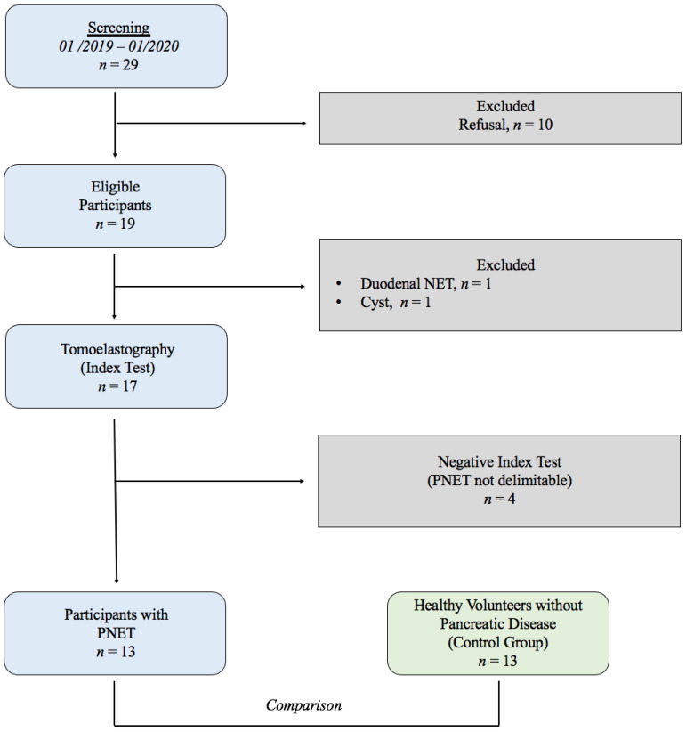
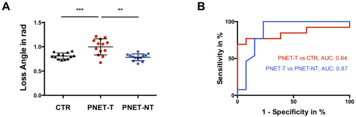
13 patients with PNET were prospectively compared to 13 age-/sex-matched heathy volunteers (CTR). Multifrequency MR elastography was combined with tomoelastography-postprocessing to provide high-resolution maps of shear wave speed (SWS in m/s). SWS of pancreatic neuroendocrine tumor (PNET-T) were compared with nontumorous pancreatic tissue in patients with PNET (PNET-NT) and heathy pancreatic tissue (CTR). The diagnostic performance of tomoelastography was evaluated by ROC-AUC analysis. PNET-SWS correlations were calculated with Pearson's .
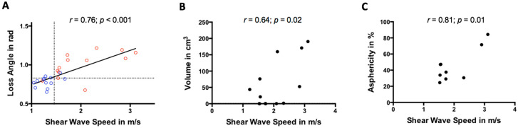
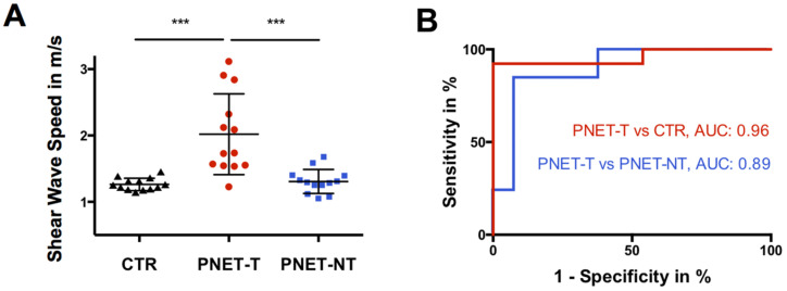
SWS was higher in PNET-T (2.02 ± 0.61 m/s) compared to PNET-NT (1.31 ± 0.18 m/s, < 0.01) and CTR (1.26 ± 0.09 m/s, < 0.01). An SWS-cutoff of 1.46 m/s distinguished PNET-T from PNET-NT (AUC = 0.89; sensitivity = 0.85; specificity = 0.92) and a cutoff of 1.49 m/s differentiated pancreatic tissue of CTR from PNET-T (AUC = 0.96; sensitivity = 0.92; specificity = 1.00). The SWS of PNET-T was positively correlated with PET derived asphericity ( = 0.81; = 0.01).
Tomoelastography provides quantitative imaging markers for the detection of PNET and the prediction of greater tumor aggressiveness by increased stiffness.
评估弹性成像断层扫描在鉴别胰腺神经内分泌肿瘤(PNET)与健康胰腺组织方面的诊断性能,并通过将PNET硬度与PET衍生的非球形度相关联来评估肿瘤侵袭性的预测。
前瞻性地将13例PNET患者与13例年龄和性别匹配的健康志愿者(对照组)进行比较。多频磁共振弹性成像与弹性成像断层扫描后处理相结合,以提供剪切波速度(SWS,单位为m/s)的高分辨率图。将PNET患者的胰腺神经内分泌肿瘤(PNET-T)的SWS与非肿瘤性胰腺组织(PNET-NT)和健康胰腺组织(对照组)进行比较。通过ROC-AUC分析评估弹性成像断层扫描的诊断性能。用Pearson相关性分析计算PNET-SWS的相关性。
与PNET-NT(1.31±0.18 m/s,P<0.01)和对照组(1.26±0.09 m/s,P<0.01)相比,PNET-T的SWS更高(2.02±0.61 m/s)。SWS临界值为1.46 m/s可区分PNET-T与PNET-NT(AUC=0.89;敏感性=0.85;特异性=0.92),临界值为1.49 m/s可区分对照组的胰腺组织与PNET-T(AUC=0.96;敏感性=0.92;特异性=1.00)。PNET-T的SWS与PET衍生的非球形度呈正相关(r=0.81;P=0.01)。
弹性成像断层扫描为PNET的检测以及通过硬度增加预测更高的肿瘤侵袭性提供了定量成像标志物。