Hu Jiaxi, Guo Jing, Pei Yigang, Hu Ping, Li Mengsi, Sack Ingolf, Li Wenzheng
Department of Radiology, Xiangya Hospital, Central South University, Changsha, China.
Department of Radiology, Charité - Universitätsmedizin Berlin, Berlin, Germany.
Front Oncol. 2021 Aug 13;11:701336. doi: 10.3389/fonc.2021.701336. eCollection 2021.
To investigate the significance of collagen in predicting the aggressiveness of rectal tumors in patients, examined based on tomoelastography quantified stiffness and by histologically measured collagen volume fraction (CVF).
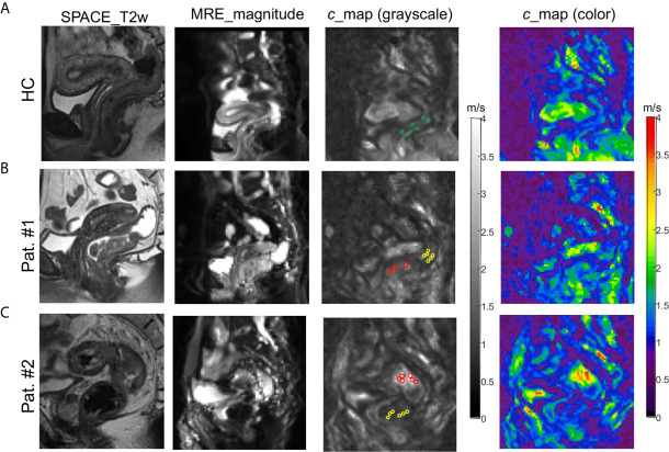
170 patients with suspected rectal cancer were prospectively enrolled and underwent preoperative magnetic resonance imaging (MRI) and rectal tomoelastography, a technique based on multifrequency magnetic resonance elastography. Histopathologic analysis identified eighty patients with rectal cancer who were divided into subgroups by tumor-node (TN) stage, prognostic stage, and risk level. Rectal tumor stiffness was correlated with histopathologic CVF. Area-under-the-curve (AUC) and contingency analysis were used to evaluate the performance of rectal stiffness in distinguishing tumor stages which was compared to standard clinical MRI.
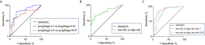
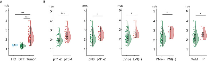
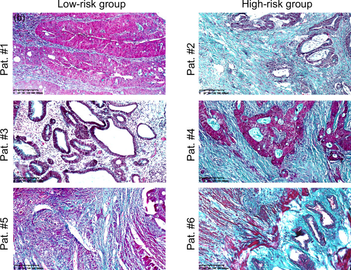
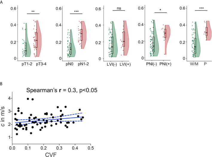
tomoelastography revealed that rectal tumor stiffened significantly with increased TN stage (p<0.05). Tumors with poorly differentiated status, perineural and lymphovascular invasion also displayed higher stiffness than well-to-moderately differentiated, noninvasive tumors (all p<0.05). Similar to stiffness, CVF indicated an abnormally high collagen content in tumors with perineural invasion and poor differentiation status. CVF was also positively correlated with stiffness (p<0.05). Most importantly, both stiffness (AUROC: 0.82) and CVF (AUROC: 0.89) demonstrated very good diagnostic accuracy in detecting rectal tumors that have high risk for progressing to an aggressive state with poorer prognosis.
In human rectal carcinomas, overexpression of collagen is correlated with increased tissue stiffness and high risk for tumor advancing more aggressively. tomoelastography quantifies rectal tumor stiffness which improves the diagnostic performance of standard MRI in the assessment of lymph nodes metastasis. Therefore, stiffness mapping by tomoelastography can predict rectal tumor aggressiveness and add diagnostic value to MRI.
基于弹性成像定量刚度和组织学测量的胶原体积分数(CVF),研究胶原在预测直肠癌患者肿瘤侵袭性中的意义。
前瞻性纳入170例疑似直肠癌患者,术前行磁共振成像(MRI)和直肠弹性成像检查,后者是一种基于多频磁共振弹性成像的技术。组织病理学分析确定了80例直肠癌患者,并根据肿瘤-淋巴结(TN)分期、预后分期和风险水平将其分为亚组。直肠肿瘤硬度与组织病理学CVF相关。采用曲线下面积(AUC)和列联分析评估直肠硬度在区分肿瘤分期方面的性能,并与标准临床MRI进行比较。
弹性成像显示,直肠肿瘤硬度随TN分期增加而显著增加(p<0.05)。低分化、神经周围和脉管侵犯的肿瘤也比高-中分化、无侵犯的肿瘤表现出更高的硬度(均p<0.05)。与硬度相似,CVF表明神经周围侵犯和低分化状态的肿瘤中胶原含量异常高。CVF也与硬度呈正相关(p<0.05)。最重要的是,硬度(AUROC:0.82)和CVF(AUROC:0.89)在检测有进展为侵袭性状态且预后较差高风险的直肠肿瘤方面均显示出非常好的诊断准确性。
在人类直肠癌中,胶原的过表达与组织硬度增加和肿瘤更具侵袭性进展的高风险相关。弹性成像可定量直肠肿瘤硬度,提高标准MRI在评估淋巴结转移中的诊断性能。因此,弹性成像的硬度图可预测直肠肿瘤的侵袭性,并为MRI增加诊断价值。