Hou Yufeng, Bai Jizhong, Shen Xin, de Langen Oscar, Li Amy, Lal Sean, Dos Remedios Cristobal G, Baddeley David, Ruygrok Peter N, Soeller Christian, Crossman David J
Department of Physiology, University of Auckland, Auckland, New Zealand.
Institute for Experimental Medical Research, Oslo University Hospital, University of Oslo, Oslo, Norway.
Front Physiol. 2021 Oct 8;12:724372. doi: 10.3389/fphys.2021.724372. eCollection 2021.
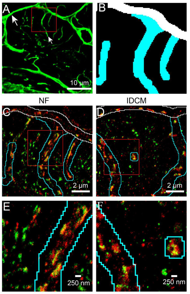
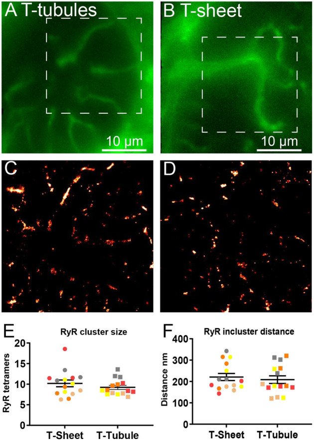
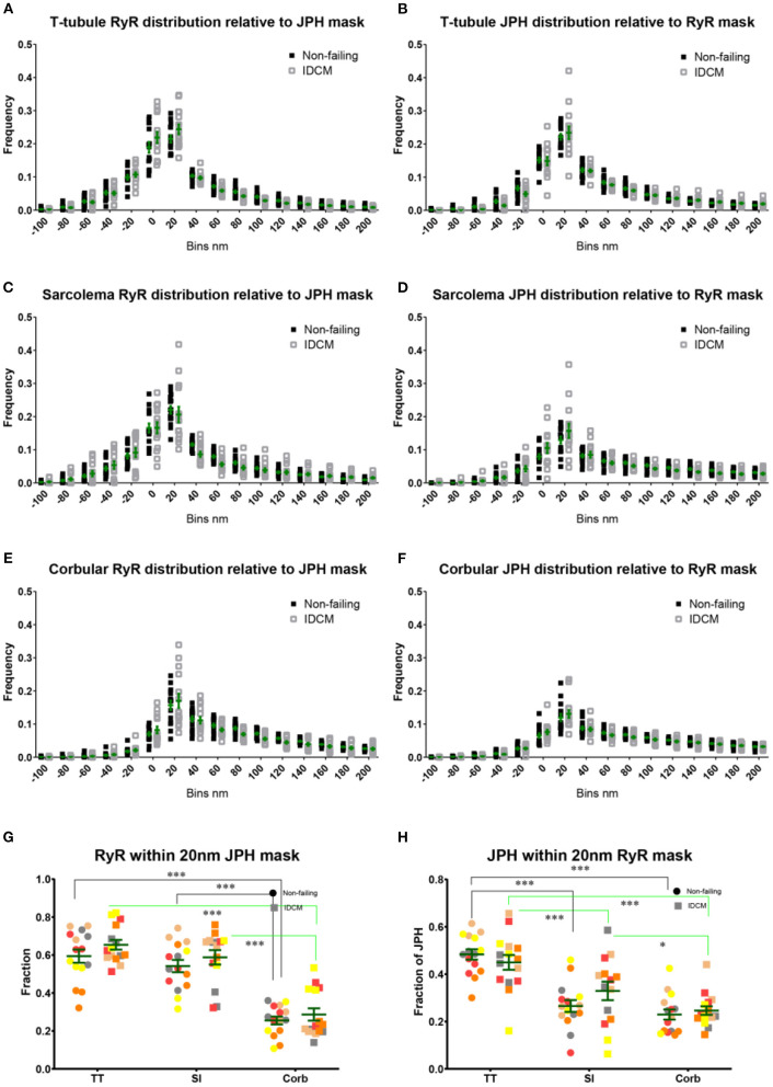
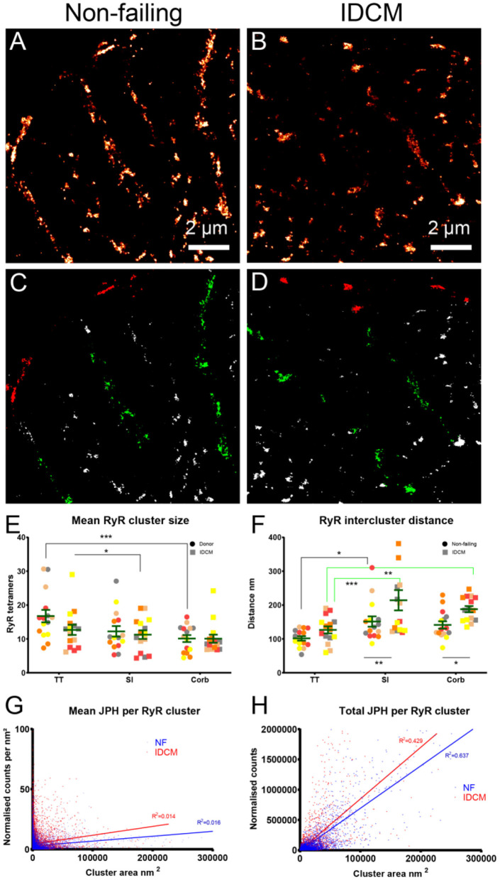
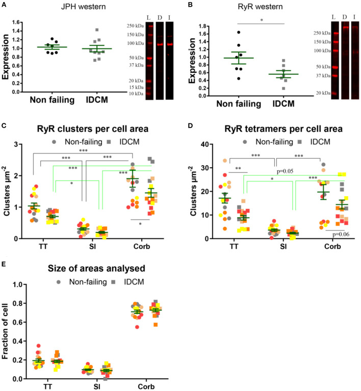
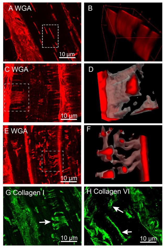
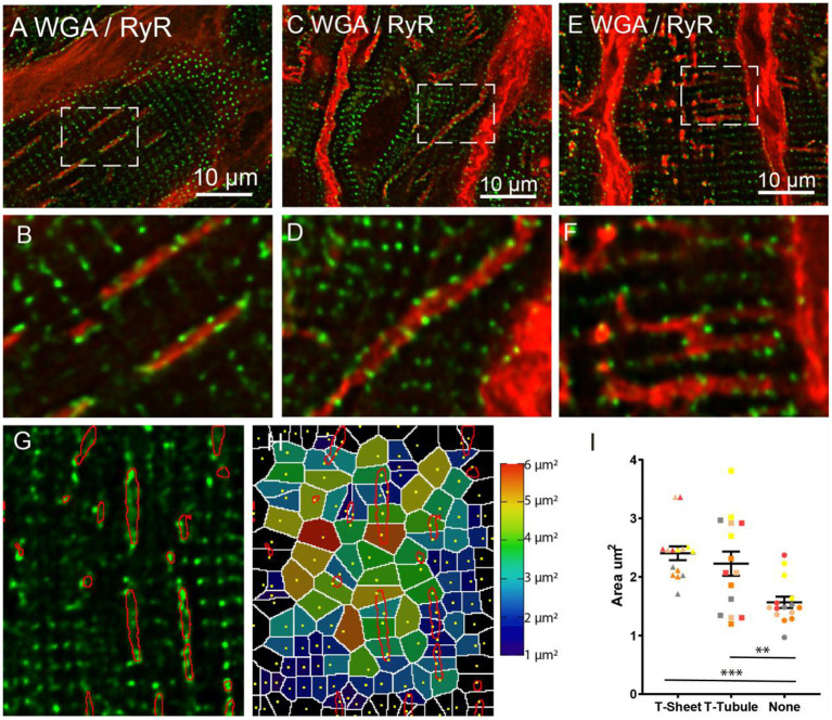
The disrupted organisation of the ryanodine receptors (RyR) and junctophilin (JPH) is thought to underpin the transverse tubule (t-tubule) remodelling in a failing heart. Here, we assessed the nanoscale organisation of these two key proteins in the failing human heart. Recently, an advanced feature of the t-tubule remodelling identified large flattened t-tubules called t-sheets, that were several microns wide. Previously, we reported that in the failing heart, the dilated t-tubules up to 1 μm wide had increased collagen, and we hypothesised that the t-sheets would also be associated with collagen deposits. Direct stochastic optical reconstruction microscopy (dSTORM), confocal microscopy, and western blotting were used to evaluate the cellular distribution of excitation-contraction structures in the cardiac myocytes from patients with idiopathic dilated cardiomyopathy (IDCM) compared to myocytes from the non-failing (NF) human heart. The dSTORM imaging of RyR and JPH found no difference in the colocalisation between IDCM and NF myocytes, but there was a higher colocalisation at the t-tubule and sarcolemma compared to the corbular regions. Western blots revealed no change in the JPH expression but did identify a ~50% downregulation of RyR ( = 0.02). The dSTORM imaging revealed a trend for the smaller t-tubular RyR clusters (24%) and reduced the t-tubular RyR cluster density (~35%) that resulted in a 50% reduction of t-tubular RyR tetramers in the IDCM myocytes ( < 0.01). Confocal microscopy identified the t-sheets in all the IDCM hearts examined and found that they are associated with the reticular collagen fibres within the lumen. However, the size and density of the RyR clusters were similar in the myocyte regions associated with t-sheets and t-tubules. T-tubule remodelling is associated with a reduced RyR expression that may contribute to the reduced excitation-contraction coupling in the failing human heart.
人们认为,兰尼碱受体(RyR)和连接蛋白(JPH)的组织结构破坏是导致衰竭心脏中横小管(t小管)重塑的原因。在此,我们评估了这两种关键蛋白在衰竭人类心脏中的纳米级组织结构。最近,t小管重塑的一个先进特征是发现了称为t片层的大的扁平t小管,其宽度达数微米。此前,我们报道在衰竭心脏中,直径达约1μm的扩张t小管中胶原蛋白增加,并且我们推测t片层也会与胶原蛋白沉积有关。与来自非衰竭(NF)人类心脏的心肌细胞相比,我们使用直接随机光学重建显微镜(dSTORM)、共聚焦显微镜和蛋白质免疫印迹法来评估特发性扩张型心肌病(IDCM)患者心肌细胞中兴奋 - 收缩结构的细胞分布。对RyR和JPH的dSTORM成像发现,IDCM和NF心肌细胞之间的共定位没有差异,但与肌小节区域相比,在t小管和肌膜处有更高的共定位。蛋白质免疫印迹显示JPH表达没有变化,但确实发现RyR下调了约50%(P = 0.02)。dSTORM成像显示较小的t小管RyR簇有减少趋势(约24%),并且t小管RyR簇密度降低(约35%),这导致IDCM心肌细胞中t小管RyR四聚体减少了50%(P < 0.01)。共聚焦显微镜在所有检查的IDCM心脏中都识别出了t片层,并发现它们与管腔内的网状胶原纤维有关。然而,与t片层和t小管相关的心肌细胞区域中RyR簇的大小和密度相似。t小管重塑与RyR表达降低有关,这可能导致衰竭人类心脏中兴奋 - 收缩偶联减少。