Fiegle Dominik J, Schöber Martin, Dittrich Sven, Cesnjevar Robert, Klingel Karin, Volk Tilmann, Alkassar Muhannad, Seidel Thomas
Institute of Cellular and Molecular Physiology, Friedrich-Alexander-Universität Erlangen-Nürnberg, Erlangen, Germany.
Department of Pediatric Cardiology, Friedrich-Alexander-Universität Erlangen-Nürnberg, Erlangen, Germany.
Front Cardiovasc Med. 2021 Jan 18;7:624776. doi: 10.3389/fcvm.2020.624776. eCollection 2020.
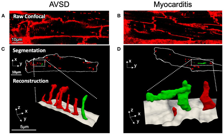
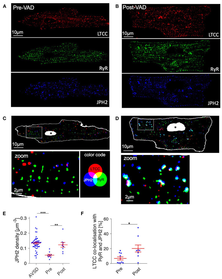
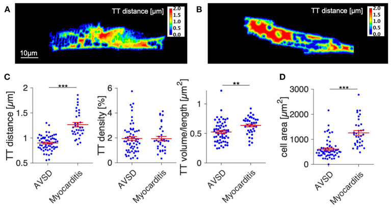
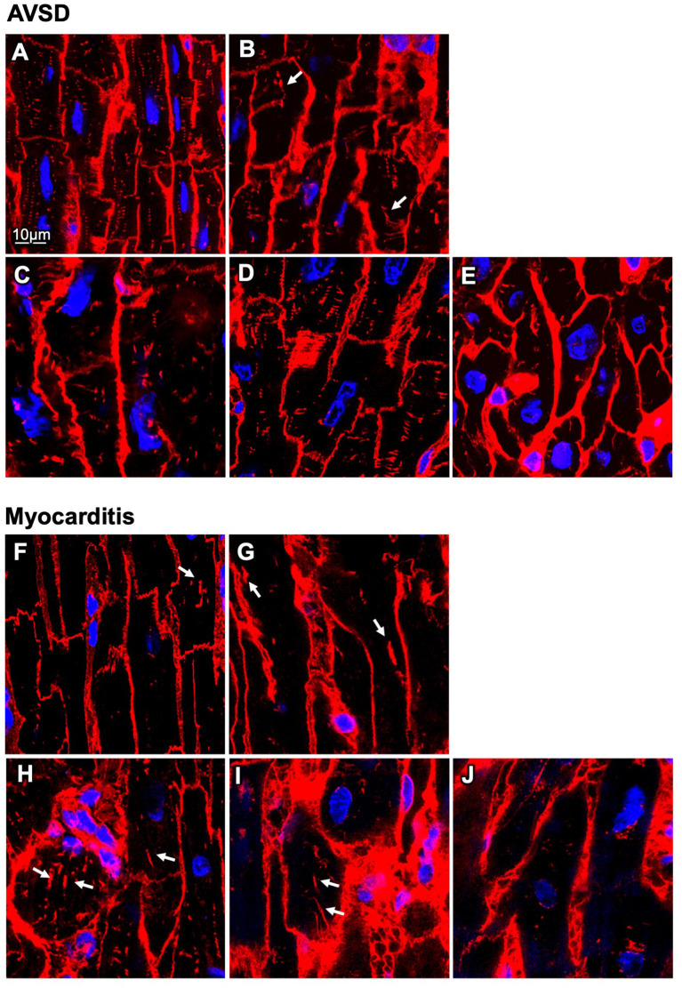
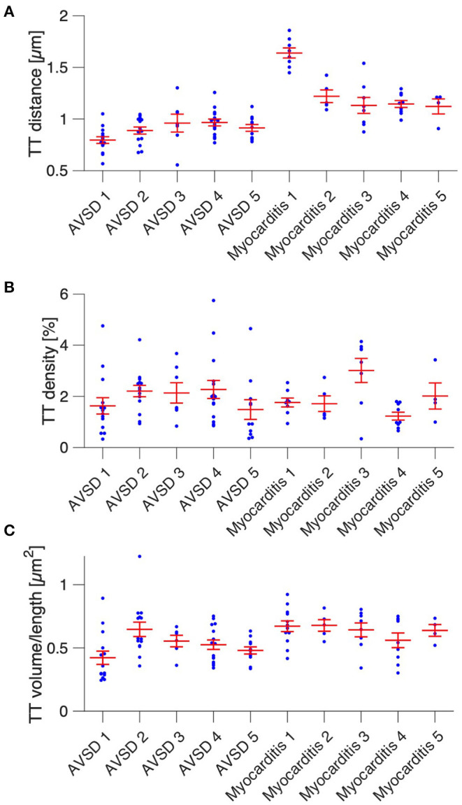
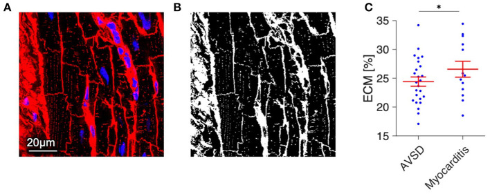
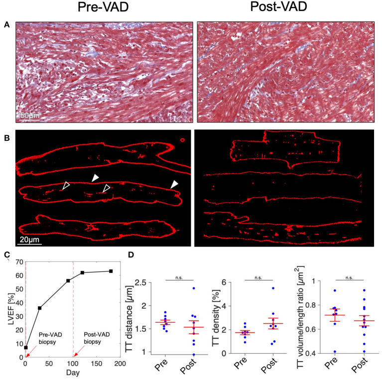
Chronic heart failure (HF) in adults causes remodeling of the cardiomyocyte transverse tubular system (t-system), which contributes to disease progression by impairing excitation-contraction (EC) coupling. However, it is unknown if t-system remodeling occurs in pediatric heart failure. This study investigated the t-system in pediatric viral myocarditis. The t-system and integrity of EC coupling junctions (co-localization of L-type Ca channels with ryanodine receptors and junctophilin-2) were analyzed by 3D confocal microscopy in left-ventricular (LV) samples from 5 children with myocarditis (age 14 ± 3 months), undergoing ventricular assist device (VAD) implantation, and 5 children with atrioventricular septum defect (AVSD, age 17 ± 3 months), undergoing corrective surgery. LV ejection fraction (EF) was 58.4 ± 2.3% in AVSD and 12.2 ± 2.4% in acute myocarditis. Cardiomyocytes from myocarditis samples showed increased t-tubule distance (1.27 ± 0.05 μm, = 34 cells) and dilation of t-tubules (volume-length ratio: 0.64 ± 0.02 μm) when compared with AVSD (0.90 ± 0.02 μm, < 0.001; 0.52 ± 0.02 μm, = 61, < 0.01). Intriguingly, 4 out of 5 myocarditis samples exhibited sheet-like t-tubules (t-sheets), a characteristic feature of adult chronic heart failure. The fraction of extracellular matrix was slightly higher in myocarditis (26.6 ± 1.4%) than in AVSD samples (24.4 ± 0.8%, < 0.05). In one case of myocarditis, a second biopsy was taken and analyzed at VAD explantation after extensive cardiac recovery (EF from 7 to 56%) and clinical remission. When compared with pre-VAD, t-tubule distance and density were unchanged, as well as volume-length ratio (0.67 ± 0.04 μm vs. 0.72 ± 0.05 μm, = 0.5), reflecting extant t-sheets. However, junctophilin-2 cluster density was considerably higher (0.12 ± 0.02 μm vs. 0.05 ± 0.01 μm, = 9/10, < 0.001), approaching values of AVSD (0.13 ± 0.05 μm, = 56), and the measure of intact EC coupling junctions showed a distinct increase (20.2 ± 5.0% vs. 6.8 ± 2.2%, < 0.001). Severe t-system loss and remodeling to t-sheets can occur in acute HF in young children, resembling the structural changes of chronically failing adult hearts. T-system remodeling might contribute to cardiac dysfunction in viral myocarditis. Although t-system recovery remains elusive, recovery of EC coupling junctions may be possible and deserves further investigation.
成人慢性心力衰竭(HF)会导致心肌细胞横管系统(t系统)重塑,通过损害兴奋-收缩(EC)偶联促进疾病进展。然而,小儿心力衰竭是否会发生t系统重塑尚不清楚。本研究调查了小儿病毒性心肌炎中的t系统。通过三维共聚焦显微镜分析了5例患有心肌炎(年龄14±3个月)并接受心室辅助装置(VAD)植入的儿童以及5例患有房室间隔缺损(AVSD,年龄17±3个月)并接受矫正手术的儿童的左心室(LV)样本中的t系统和EC偶联连接的完整性(L型钙通道与兰尼碱受体和连接蛋白-2的共定位)。AVSD组左心室射血分数(EF)为58.4±2.3%,急性心肌炎组为12.2±2.4%。与AVSD组相比,心肌炎样本中的心肌细胞显示t小管间距增加(1.27±0.05μm,n = 34个细胞)以及t小管扩张(体积-长度比:0.64±0.02μm)(AVSD组为0.90±0.02μm,P < 0.001;0.52±0.02μm,n = 61,P < 0.01)。有趣的是,5例心肌炎样本中有4例表现出片状t小管(t片层),这是成人慢性心力衰竭的一个特征性表现。心肌炎样本中细胞外基质的比例略高于AVSD样本(26.6±1.4% 对24.4±0.8%,P < 0.05)。在1例心肌炎病例中,在心脏广泛恢复(EF从7%提高到56%)且临床缓解后,于VAD取出时进行了第二次活检并分析。与VAD植入前相比,t小管间距和密度以及体积-长度比均未改变(0.67±0.04μm对0.72±0.05μm,P = 0.5),反映出t片层仍然存在。然而,连接蛋白-2簇密度显著更高(0.12±0.02μm对0.05±0.01μm,n = 9/10,P < 0.001),接近AVSD组的值(0.13±0.05μm,n = 56),并且完整EC偶联连接的测量显示有明显增加(20.2±5.0%对6.8±2.2%,P < 0.001)。幼儿急性心力衰竭时可能会发生严重的t系统丢失并重塑为t片层,类似于慢性衰竭成人心脏的结构变化。t系统重塑可能导致病毒性心肌炎中的心脏功能障碍。虽然t系统的恢复仍然难以实现,但EC偶联连接的恢复可能是可行的,值得进一步研究。