Javad Farhangi Mohammad, Es-Haghi Ali, Taghavizadeh Yazdi Mohammad Ehsan, Rahdar Abbas, Baino Francesco
Department of Biology, Mashhad Branch, Islamic Azad University, Mashhad 91871-47578, Iran.
Applied Biomedical Research Center, Mashhad University of Medical Sciences, Mashhad 91388-13944, Iran.
J Funct Biomater. 2021 Sep 28;12(4):53. doi: 10.3390/jfb12040053.
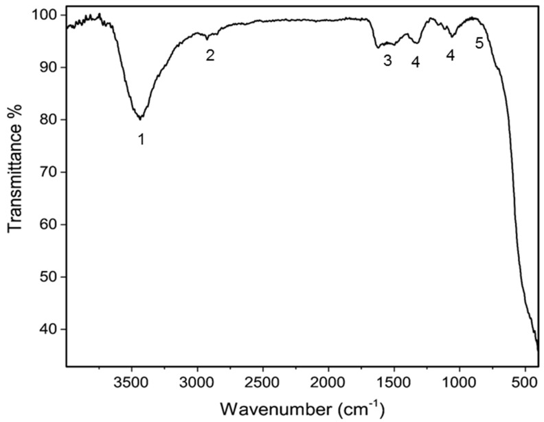
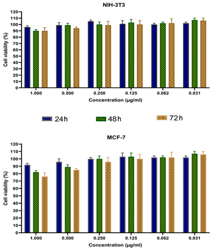
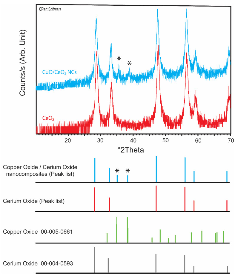
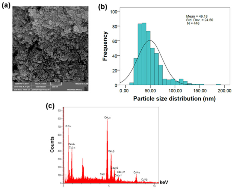
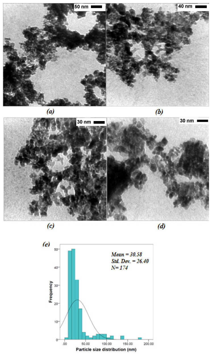
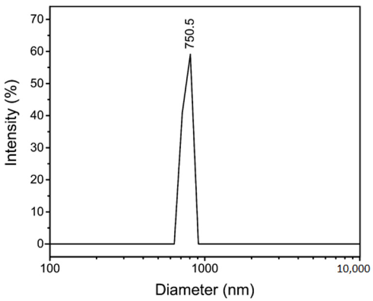
A copper oxide/cerium oxide nanocomposite (CuO/CeO, NC) was synthesized via a novel method using a metal-organic framework as a precursor. This nanomaterial was characterized by Fourier transform infrared spectroscopy (FTIR), powder X-ray diffraction (PXRD), field emission scanning electron microscopy (FESEM), transmission electron microscopy (TEM), dynamic light scattering size analysis (DLS), and zeta potential. The PXRD showed the successful synthesis of the CuO/CeO NC, in which the 2theta values of 35.55° (d = 2.52 Å, 100%) and 38.73° (d = 2.32 Å, 96%) revealed the existence of copper (II) oxide. FTIR analysis showed the CeO, hydroxyl groups, absorbed water, and some residual peaks. The solid phase analysis by FESEM and TEM images showed mean particle sizes of 49.18 ± 24.50 nm and 30.58 ± 26.40 nm, respectively, which were comparable with crystallite size (38.4 nm) obtained from PXRD, but it appears the CuO/CeO NC was not evenly distributed and in some areas, showed it was highly agglomerated. The hydrodynamic size (750.5 nm) also showed the agglomeration of the CuO/CeO NCs in the solution, which had a negatively charged surface. The CuO/CeO NCs showed anti-proliferative activity against human breast cancer cell line (MCF-7) in a dose- and time-dependence way, while affecting normal cells less significantly.
采用一种以金属有机框架为前驱体的新方法合成了氧化铜/氧化铈纳米复合材料(CuO/CeO,NC)。通过傅里叶变换红外光谱(FTIR)、粉末X射线衍射(PXRD)、场发射扫描电子显微镜(FESEM)、透射电子显微镜(TEM)、动态光散射尺寸分析(DLS)和zeta电位对该纳米材料进行了表征。PXRD表明成功合成了CuO/CeO NC,其中35.55°(d = 2.52 Å,100%)和38.73°(d = 2.32 Å,96%)的2θ值表明存在氧化铜(II)。FTIR分析显示了CeO、羟基、吸附水和一些残留峰。FESEM和TEM图像的固相分析表明,平均粒径分别为49.18±24.50 nm和30.58±26.40 nm,这与PXRD获得的微晶尺寸(38.4 nm)相当,但CuO/CeO NC似乎分布不均匀,在某些区域显示出高度团聚。流体动力学尺寸(750.5 nm)也表明CuO/CeO NC在溶液中发生了团聚,其表面带负电荷。CuO/CeO NC对人乳腺癌细胞系(MCF-7)具有剂量和时间依赖性的抗增殖活性,而对正常细胞的影响较小。