Department of Anesthesiology, Affiliated Baoan Central Hospital of Guangdong Medical University, No 60 Leyuan Road, Baoan Distric of Shenzhen, Shenzhen, Guangdong Province, China.
Department of Anesthesiology, Sichuan Academy of Medical Science & Sichuan Provincial People's Hospital, Electronic Science and Technology University, 18 Huanhua Road, Chengdu, China.
BMC Anesthesiol. 2021 Oct 26;21(1):257. doi: 10.1186/s12871-021-01475-7.
Angiogenesis, the formation of blood vessel from pre-existing ones, plays an important role in many pathophysiological diseases, such as cancer. Opioids are often used in clinic for the management of chronic pain in cancer patients at terminal phases. Here, we investigated and compared the effects and mechanisms of four opioids on angiogenesis.
We performed angiogenesis assays on human umbilical vein endothelial cells (HUVEC) that represent an in vitro model to assess the toxicity of drugs to endothelium.
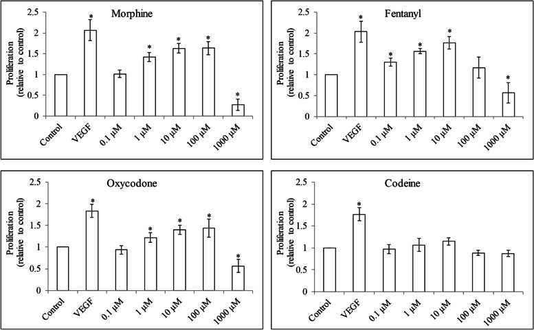
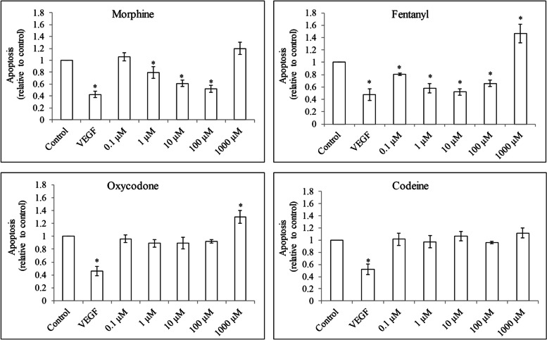
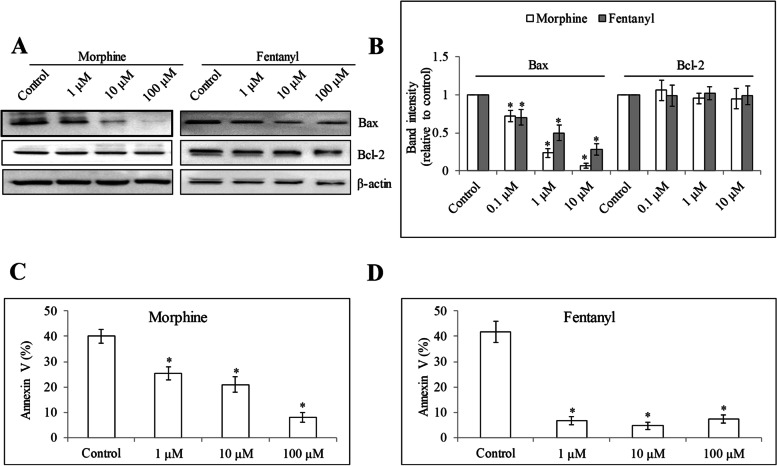
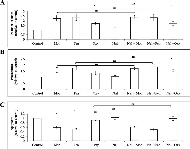
Morphine and oxycodone at 0.1 μM to 100 μM dose-dependently increased endothelial cell tube formation and proliferation. We observed the same in endothelial cells exposed to fentanyl at 0.1 μM to 10 μM but there was a gradual loss of stimulation by fentanyl at 100 μM and 1000 μM. Morphine and fentanyl reduced endothelial cell apoptosis-induced by serum withdrawal whereas oxycodone did not display anti-apoptotic effect, via decreasing Bax level. Oxycodone at the same concentrations was less potent than morphine and fentanyl. Different from other three opioids, codeine at all tested concentrations did not affect endothelial cell tube formation, proliferation and survival. Mechanism studies demonstrated that opioids acted on endothelial cells via μ-opioid receptor-independent pathway. Although we observed the increased phosphorylation of mitogen-activated protein kinase (MAPK) in cells exposed to morphine, fentanyl and oxycodone, the rescue studies demonstrated that the stimulatory effects of morphine but not fentanyl nor oxycodone were reversed by a specific MAPK inhibitor.
Our work demonstrates the differential effects and mechanisms of opioids on angiogenesis.
血管生成,即从预先存在的血管中形成新的血管,在许多病理生理疾病中起着重要作用,如癌症。阿片类药物常用于临床治疗癌症终末期患者的慢性疼痛。在这里,我们研究并比较了四种阿片类药物对血管生成的影响及其机制。
我们在人脐静脉内皮细胞(HUVEC)上进行了血管生成实验,该细胞是评估药物对内皮细胞毒性的体外模型。
吗啡和羟考酮在 0.1μM 至 100μM 的剂量范围内,呈剂量依赖性地增加内皮细胞管状结构形成和增殖。我们在暴露于 0.1μM 至 10μM 芬太尼的内皮细胞中观察到了同样的情况,但在 100μM 和 1000μM 时,芬太尼的刺激作用逐渐丧失。吗啡和芬太尼减少了因血清去除而导致的内皮细胞凋亡,而羟考酮则通过降低 Bax 水平没有显示出抗凋亡作用。在相同浓度下,羟考酮的作用不及吗啡和芬太尼。与其他三种阿片类药物不同,在所有测试浓度下,可待因均不影响内皮细胞管状结构的形成、增殖和存活。机制研究表明,阿片类药物通过μ-阿片受体非依赖性途径作用于内皮细胞。尽管我们观察到暴露于吗啡、芬太尼和羟考酮的细胞中丝裂原活化蛋白激酶(MAPK)的磷酸化增加,但挽救研究表明,吗啡的刺激作用而非芬太尼或羟考酮的刺激作用可被特定的 MAPK 抑制剂逆转。
我们的工作表明阿片类药物对血管生成的影响及其机制存在差异。