Pulmonary & Critical Care Medicine Division, Department of Internal Medicine, University of Michigan Medical School and Michigan Medicine, Ann Arbor, Michigan.
Graduate Program in Immunology, University of Michigan, Ann Arbor, Michigan.
Am J Physiol Lung Cell Mol Physiol. 2021 Dec 1;321(6):L1183-L1193. doi: 10.1152/ajplung.00322.2020. Epub 2021 Oct 27.
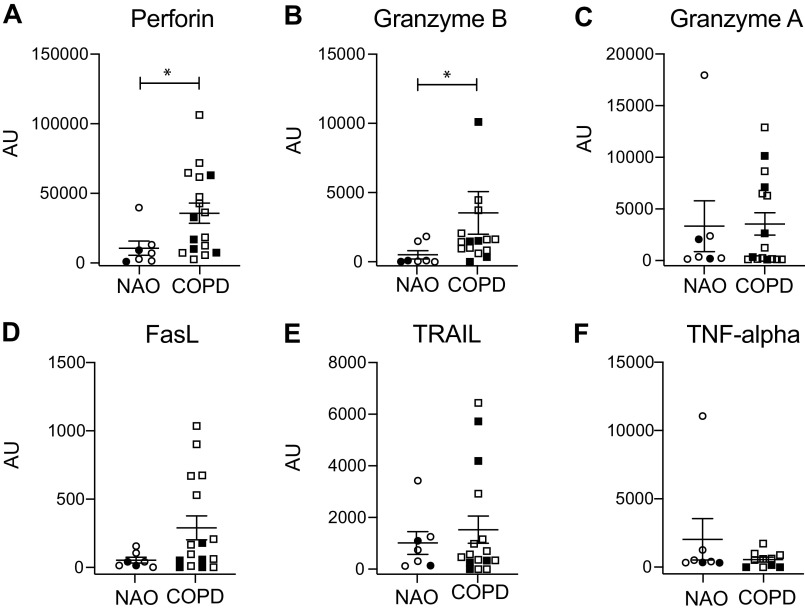
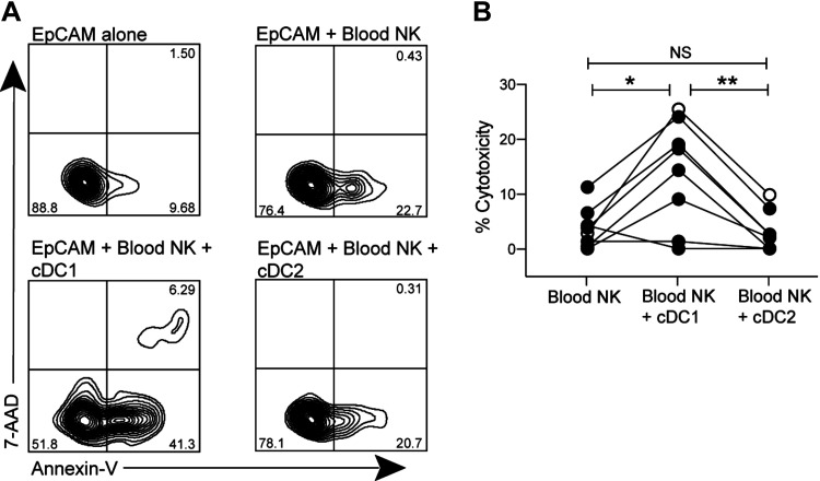
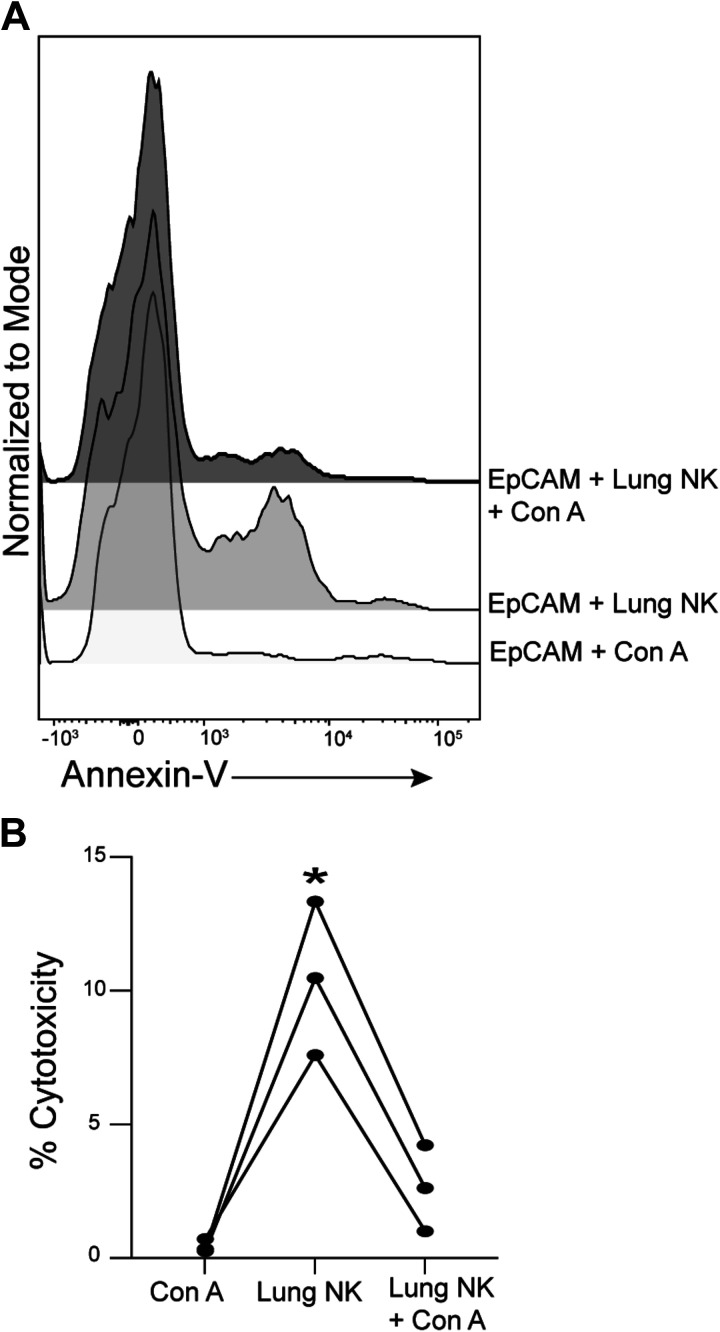
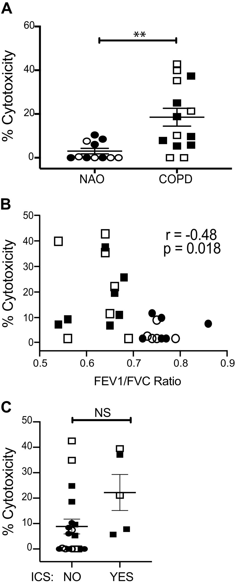
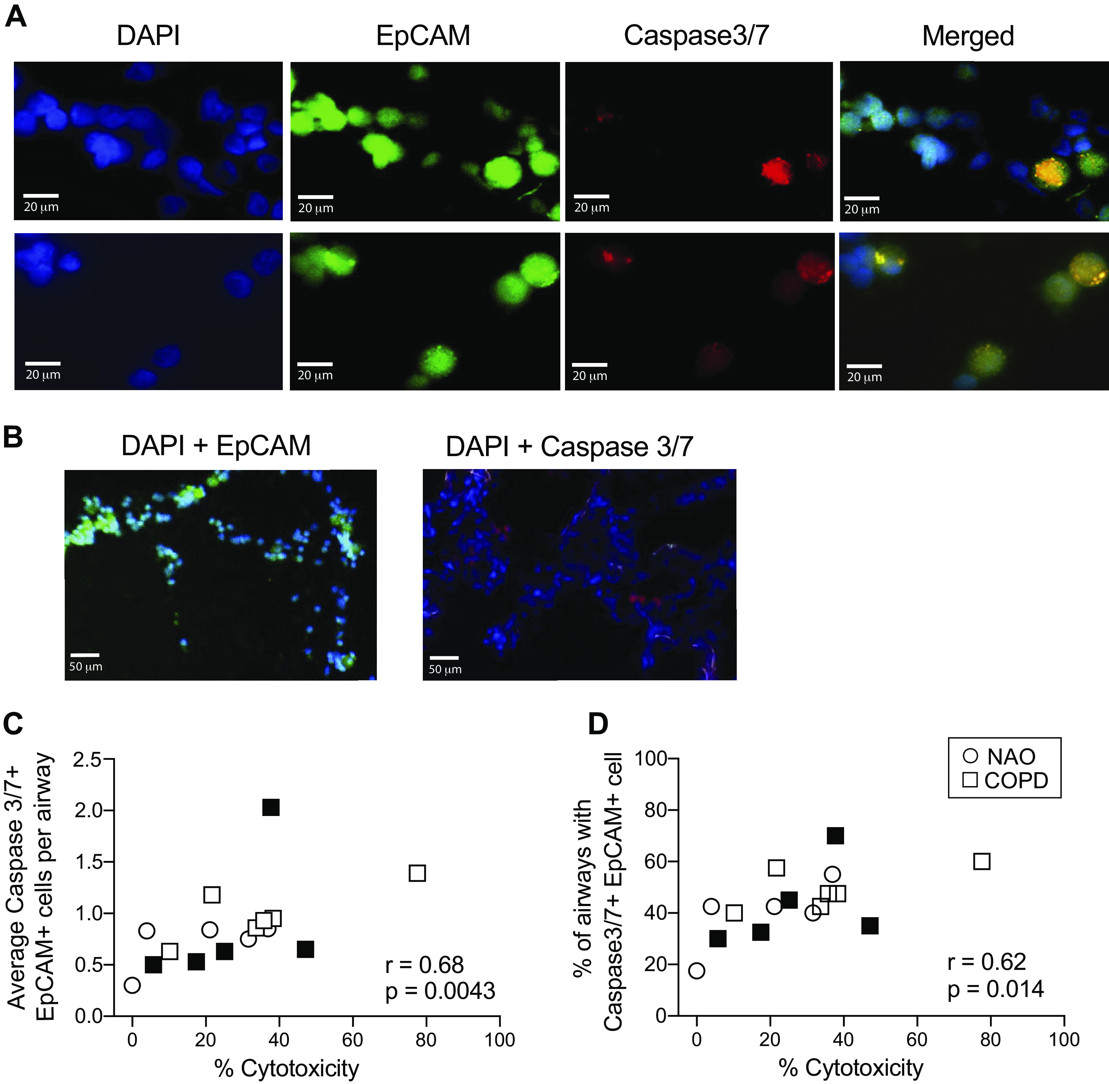
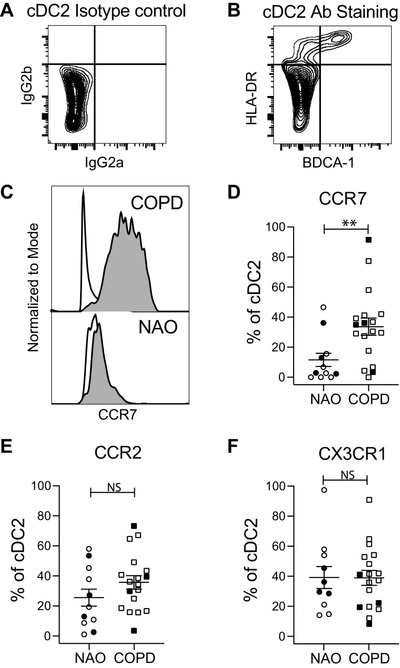
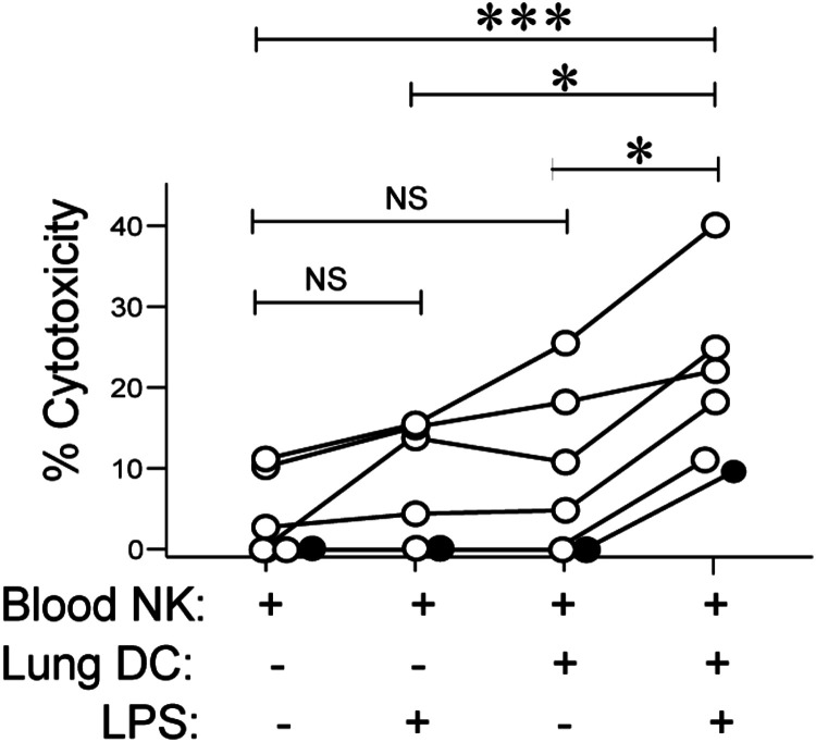
In chronic obstructive pulmonary disease (COPD), lung natural killer cells (NKs) lyse autologous lung epithelial cells in vitro, but underlying mechanisms and their relationship to epithelial cell apoptosis in vivo are undefined. Although this cytolytic capacity of lung NKs depends on priming by dendritic cells (DCs), whether priming correlates with DC maturation or is limited to a specific DC subset is also unknown. We recruited ever-smokers (≥10 pack-years; = 96) undergoing clinically indicated lung resections. We analyzed lung NKs for cytotoxic molecule transcripts and for cytotoxicity, which we correlated with in situ detection of activated Caspase-3/7+ airway epithelial cells. To investigate DC priming, we measured lung DC expression of CCR2, CCR7, and CX3CR1 and cocultured peripheral blood NKs with autologous lung DCs, either matured using lipopolysaccharide (LPS) (nonobstructed smokers) or separated into conventional dendritic cell type-1 (cDC1) versus cDC type-2 (cDC2) (COPD). Lung NKs in COPD expressed more perforin ( < 0.02) and granzyme B ( < 0.03) transcripts; inhibiting perforin blocked in vitro killing by lung NKs. Cytotoxicity in vitro correlated significantly ( = 0.68, = 0.0043) with numbers of apoptotic epithelial cells per airway. In nonobstructed smokers, LPS-induced maturation enhanced DC-mediated priming of blood NKs, reflected by greater epithelial cell death. Although CCR7 expression was greater in COPD in both cDC1 ( < 0.03) and cDC2 ( = 0.009), only lung cDC1 primed NK killing. Thus, rather than being intrinsic to those with COPD, NK priming is a capacity of human lung DCs that is inducible by recognition of bacterial (and possibly other) danger signals and restricted to the cDC1 subset.
在慢性阻塞性肺疾病(COPD)中,肺自然杀伤细胞(NK 细胞)在体外裂解自身肺上皮细胞,但潜在机制及其与体内上皮细胞凋亡的关系尚不清楚。虽然肺 NK 细胞的这种细胞溶解能力依赖于树突状细胞(DC)的启动,但启动是否与 DC 成熟相关,还是仅限于特定的 DC 亚群尚不清楚。我们招募了正在接受临床指征性肺切除术的长期吸烟者(≥10 包年;n = 96)。我们分析了肺 NK 细胞的细胞毒性分子转录物和细胞毒性,将其与原位检测到的激活 Caspase-3/7+气道上皮细胞进行了相关性分析。为了研究 DC 启动,我们测量了肺 DC 表达 CCR2、CCR7 和 CX3CR1,并将外周血 NK 细胞与自体肺 DC 共培养,这些 DC 要么使用脂多糖(LPS)成熟(非阻塞性吸烟者),要么分离为传统树突状细胞 1 型(cDC1)和 cDC2(COPD)。COPD 患者的肺 NK 细胞表达更多的穿孔素(<0.02)和颗粒酶 B(<0.03)转录物;抑制穿孔素可阻断肺 NK 细胞的体外杀伤。体外细胞毒性与每个气道的凋亡上皮细胞数量显著相关(r = 0.68,p = 0.0043)。在非阻塞性吸烟者中,LPS 诱导的成熟增强了 DC 介导的血液 NK 细胞的启动,这反映在更大的上皮细胞死亡上。尽管 CCR7 在 COPD 患者的 cDC1(<0.03)和 cDC2(p = 0.009)中表达更高,但只有肺 cDC1 可启动 NK 细胞的杀伤作用。因此,NK 细胞的启动不是 COPD 患者所特有的,而是人类肺 DC 的一种能力,这种能力可通过识别细菌(和可能其他)危险信号而被诱导,并仅限于 cDC1 亚群。