Wake Nicole, Rosenkrantz Andrew B, Huang William C, Wysock James S, Taneja Samir S, Sodickson Daniel K, Chandarana Hersh
Department of Radiology, Montefiore Medical Center, Albert Einstein College of Medicine, 111 East 210th Street, Bronx, NY, 10467, USA.
Center for Advanced Imaging Innovation and Research (CAI2R) and Bernard and Irene Schwartz Center for Biomedical Imaging, Department of Radiology, NYU Langone Health, NYU Grossman School of Medicine, New York, NY, USA.
3D Print Med. 2021 Oct 28;7(1):34. doi: 10.1186/s41205-021-00125-5.
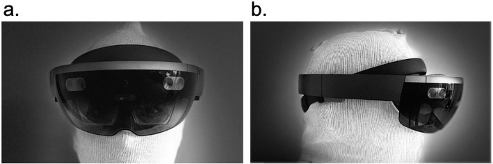
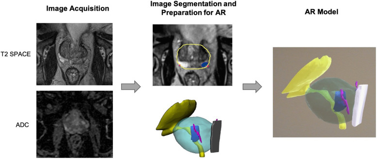
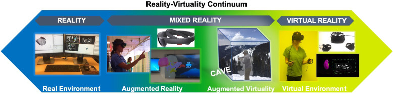
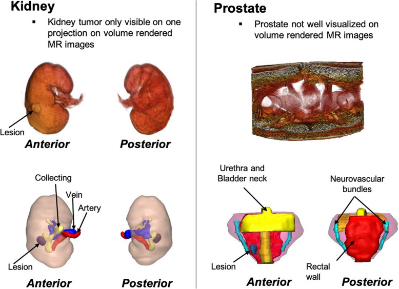
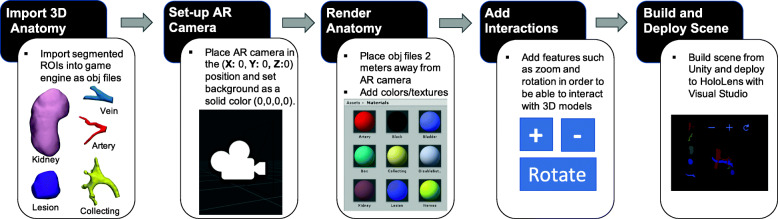
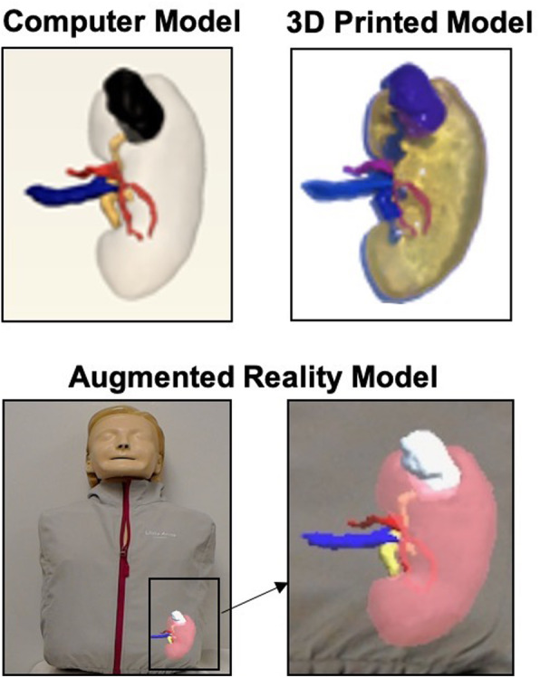
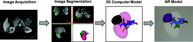
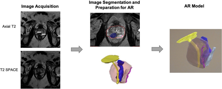
Augmented reality (AR) and virtual reality (VR) are burgeoning technologies that have the potential to greatly enhance patient care. Visualizing patient-specific three-dimensional (3D) imaging data in these enhanced virtual environments may improve surgeons' understanding of anatomy and surgical pathology, thereby allowing for improved surgical planning, superior intra-operative guidance, and ultimately improved patient care. It is important that radiologists are familiar with these technologies, especially since the number of institutions utilizing VR and AR is increasing. This article gives an overview of AR and VR and describes the workflow required to create anatomical 3D models for use in AR using the Microsoft HoloLens device. Case examples in urologic oncology (prostate cancer and renal cancer) are provided which depict how AR has been used to guide surgery at our institution.
增强现实(AR)和虚拟现实(VR)是新兴技术,有潜力极大地提升患者护理水平。在这些增强的虚拟环境中可视化特定患者的三维(3D)成像数据,可能会改善外科医生对解剖结构和手术病理学的理解,从而实现更好的手术规划、更出色的术中指导,并最终提升患者护理质量。放射科医生熟悉这些技术很重要,特别是鉴于使用VR和AR的机构数量在不断增加。本文概述了AR和VR,并描述了使用微软HoloLens设备创建用于AR的解剖3D模型所需的工作流程。提供了泌尿肿瘤学(前列腺癌和肾癌)的病例示例,展示了AR在我们机构如何用于指导手术。