Liu Shu-Ya, Liao Yin, Hosseinifard Hossein, Imani Saber, Wen Qing-Lian
Department of Oncology, The Affiliated Hospital of Southwest Medical University, Luzhou, China.
Department of Oncology, Chengdu Jinniu District People's Hospital, Chengdu, China.
Front Cell Dev Biol. 2021 Oct 15;9:705791. doi: 10.3389/fcell.2021.705791. eCollection 2021.
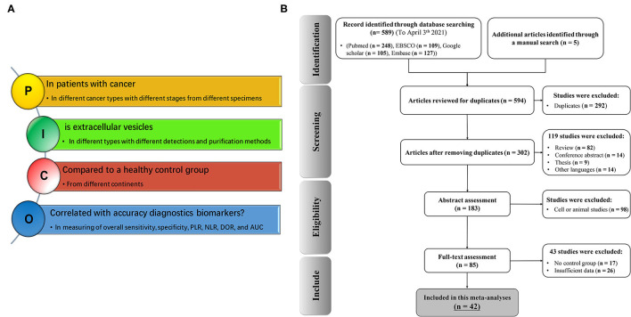
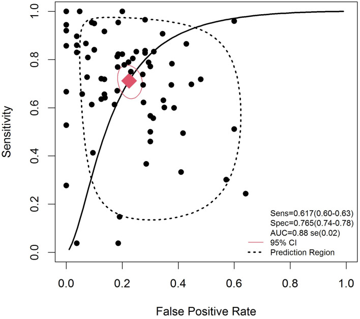
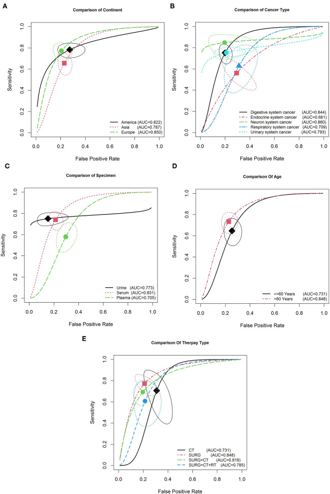
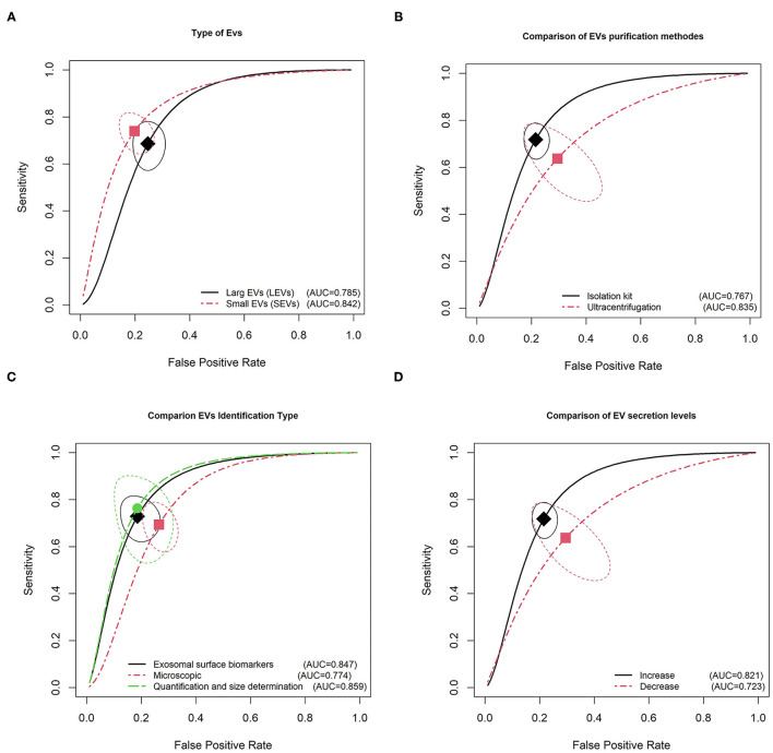
Cancer-derived extracellular vesicles (EVs) are regarded to have significant function in most steps during cancer progression. This meta-analysis aims to investigate the accuracy of EVs as a biomarker in cancer diagnosis. The diagnostic efficacy of EVs for different cancers was assessed using pooled sensitivity and specificity, diagnostic odds ratio (DOR), and overall area under the curve (AUC) of the summary receiver operating characteristic (SROC). The positive likelihood ratio (PLR) and negative likelihood ratio (NLR) were verified to estimate the diagnostic efficacy of EV at a clinical level. In all, 6,183 cancer patients and 2,437 healthy controls from 75 eligible studies reported in 42 publications were included in the study. The overall pooled sensitivity, specificity, PLR, NLR, and DOR were 0.62 (95% CI: 0.60-0.63), 0.76 (95% CI: 0.75-0.78), 3.07 (95% CI: 2.52-3.75), 0.34 (95% CI: 0.28-0.41), and 10.98 (95% CI: 7.53-16.00), respectively. Similarly, the AUC of the SROC was 0.88, indicating a high conservation of EVs as an early diagnostic marker. Furthermore, subgroup analysis suggested that the use of small EVs as a biomarker was more accurate in serum-based samples of nervous system cancer ( < 0.001). As a result, ultracentrifugation and quantification and size determination methods, such as Western blotting and ELISA were the most reliable identification methods for EV detection. We also indicated that increased secretion of EVs made them a capable biomarker for diagnosing cancer in elderly European individuals. Our study provides evidence that EVs are a promising non-invasive biomarker for cancer diagnosis. Well-designed cohort studies should be conducted to warrant the clinical diagnostic value of EVs.
癌症衍生的细胞外囊泡(EVs)被认为在癌症进展的大多数阶段都具有重要作用。本荟萃分析旨在研究EVs作为癌症诊断生物标志物的准确性。使用汇总敏感性和特异性、诊断比值比(DOR)以及汇总接受者操作特征曲线(SROC)的曲线下总面积(AUC)来评估EVs对不同癌症的诊断效能。验证了阳性似然比(PLR)和阴性似然比(NLR)以估计EVs在临床水平的诊断效能。本研究共纳入了42篇出版物中报道的75项符合条件研究的6183例癌症患者和2437例健康对照。总体汇总敏感性、特异性、PLR、NLR和DOR分别为0.62(95%CI:0.60 - 0.63)、0.76(95%CI:0.75 - 0.78)、3.07(95%CI:2.52 - 3.75)、0.34(95%CI:0.28 - 0.41)和10.98(95%CI:7.53 - 16.00)。同样,SROC的AUC为0.88,表明EVs作为早期诊断标志物具有高度的一致性。此外,亚组分析表明,在神经系统癌症的血清样本中,使用小EVs作为生物标志物更准确(<0.001)。因此,超速离心以及蛋白质免疫印迹和酶联免疫吸附测定等定量和大小测定方法是EV检测最可靠的鉴定方法。我们还指出,EVs分泌增加使其成为诊断老年欧洲个体癌症的有效生物标志物。我们的研究提供了证据表明EVs是一种有前景的癌症诊断非侵入性生物标志物。应开展设计良好的队列研究以确保EVs的临床诊断价值。