• 文献检索
  • 文档翻译
  • 深度研究
  • 学术资讯
  • Suppr Zotero 插件Zotero 插件
  • 邀请有礼
  • 套餐&价格
  • 历史记录
应用&插件
Suppr Zotero 插件Zotero 插件浏览器插件Mac 客户端Windows 客户端微信小程序
定价
高级版会员购买积分包购买API积分包
服务
文献检索文档翻译深度研究API 文档MCP 服务
关于我们
关于 Suppr公司介绍联系我们用户协议隐私条款
关注我们

Suppr 超能文献

核心技术专利:CN118964589B侵权必究
粤ICP备2023148730 号-1Suppr @ 2026

文献检索

告别复杂PubMed语法,用中文像聊天一样搜索,搜遍4000万医学文献。AI智能推荐,让科研检索更轻松。

立即免费搜索

文件翻译

保留排版,准确专业,支持PDF/Word/PPT等文件格式,支持 12+语言互译。

免费翻译文档

深度研究

AI帮你快速写综述,25分钟生成高质量综述,智能提取关键信息,辅助科研写作。

立即免费体验

实性透明细胞肾细胞癌≤4cm 的超声造影特征诊断:一项荟萃分析。

Characteristics of contrast-enhanced ultrasound for diagnosis of solid clear cell renal cell carcinomas ≤4 cm: A meta-analysis.

机构信息

Department of Ultrasound, The Affiliated Hospital of North China University of Science and Technology, Tangshan, Hebei, P.R. China.

Department of Ultrasound, Tianjin Third Central Hospital, Tianjin, P.R. China.

出版信息

Cancer Med. 2021 Dec;10(23):8288-8299. doi: 10.1002/cam4.4365. Epub 2021 Nov 1.

DOI:10.1002/cam4.4365
PMID:34725960
原文链接:https://pmc.ncbi.nlm.nih.gov/articles/PMC8633224/
Abstract

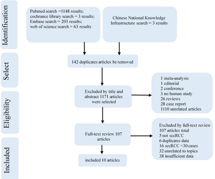
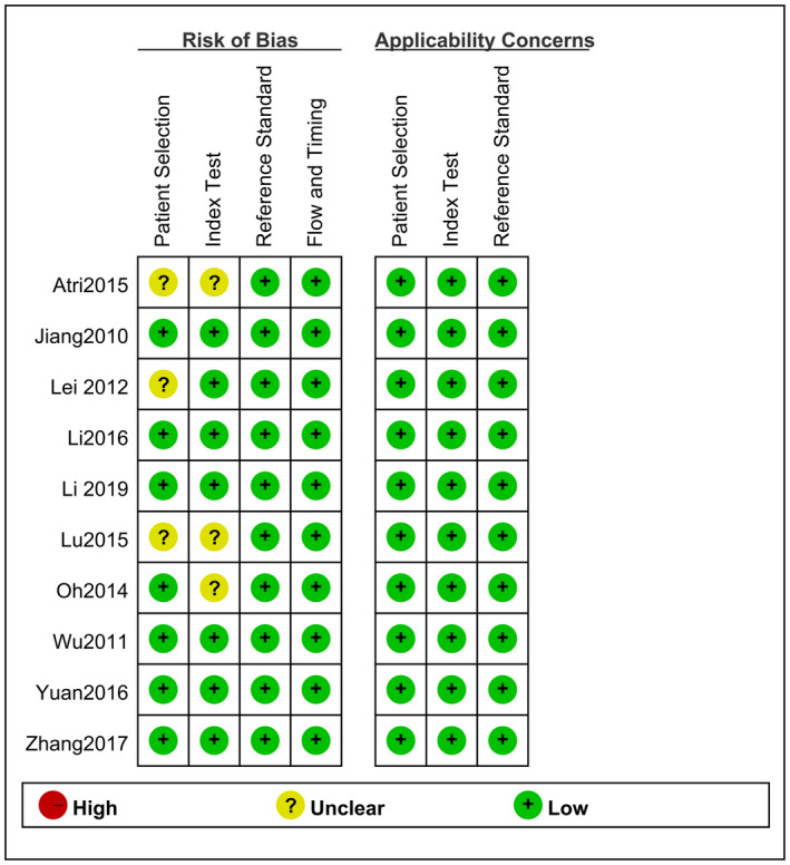
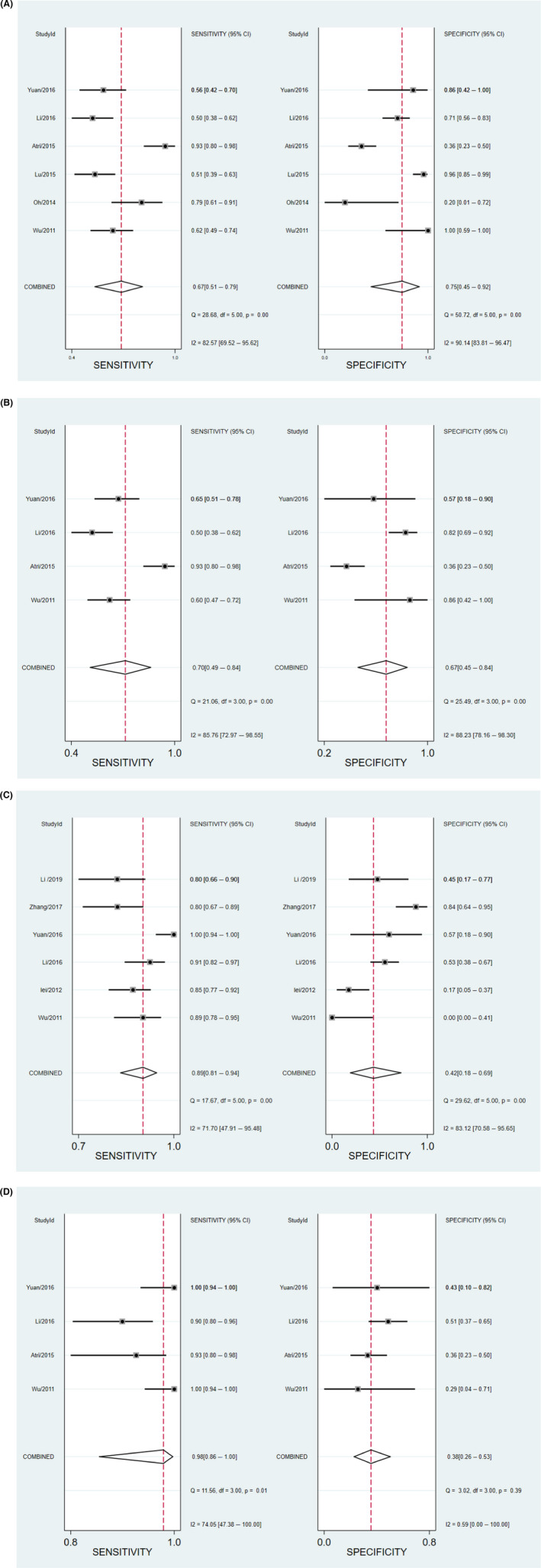
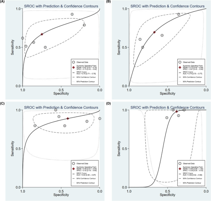
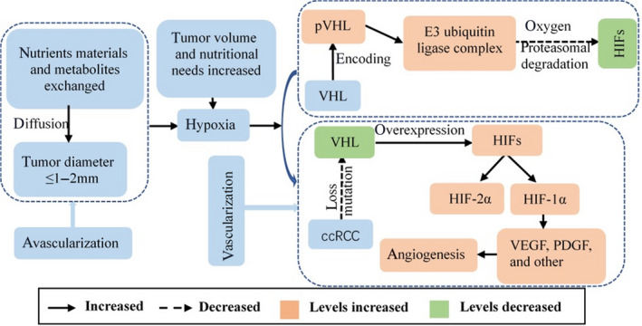
Now solid renal tumors ≤4 cm is the most common, especially the subtype of clear cell renal cell carcinoma (ccRCC) of malignant kidney tumors in clinical. However, there is not specific characteristics of contrast-enhanced ultrasound (CEUS) be recommended by the EFSUMB Guidelines in distinguish the essence of the kidney tumor with different sizes. Therefore, this meta-analysis aimed to assess the ability of CEUS to diagnose solid ccRCC (sccRCC) ≤4 cm. We comprehensively searched the Cochrane Library, Embase, PubMed, and Web of Science databases from their inception to 28 July 2020, for studies reporting the CEUS features of sccRCC lesions ≤4 cm. Additional articles were identified through the Chinese National Knowledge Infrastructure database. Studies were selected independently by two investigators and the relevant data were extracted. Discrepancies were resolved via discussion with the senior author. Study quality was assessed using the Quality Assessment of Diagnostic Accuracy Studies-2 tool, and the sensitivity and specificity of each study were determined and plotted as a receiver operating characteristic curve. Ten studies were included in this meta-analysis. Hyperenhancement showed medium sensitivity (67%-89%) and specificity (42%-75%) for diagnosing sccRCC ≤4 cm, fast-in contrast agent and heterogeneous enhancement showed high diagnostic abilities (area under curve (AUC) 0.74-0.84), but the presence of a pseudocapsule and fast-out contrast agent had poor diagnostic ability (AUC <0.70). The combination of hyperenhancement and iso-enhancement showed high sensitivity (98%) for diagnosing sccRCC ≤4 cm. Hyperenhancement, fast-in contrast agent, and heterogeneous enhancement may be specific features that could help to identify sccRCC ≤4 cm, while the presence of a pseudocapsule and fast-out of contrast agent may have low diagnostic values. The combination of multiple indexes may improve the diagnostic value of CEUS for sccRCC ≤4 cm.

摘要

目前,直径≤4cm 的肾脏实体肿瘤最为常见,尤其是临床中恶性肾肿瘤的透明细胞肾细胞癌(ccRCC)亚型。然而,EFSUMB 指南并未推荐特定的对比增强超声(CEUS)特征来区分不同大小的肾肿瘤的本质。因此,这项荟萃分析旨在评估 CEUS 诊断直径≤4cm 的肾脏实体 ccRCC(sccRCC)的能力。我们全面检索了 Cochrane 图书馆、Embase、PubMed 和 Web of Science 数据库,检索时间从建库至 2020 年 7 月 28 日,以获取报告直径≤4cm 的 sccRCC 病变的 CEUS 特征的研究。还通过中国国家知识基础设施数据库识别了其他文章。由两名研究者独立选择研究,并提取相关数据。通过与资深作者讨论解决分歧。使用诊断准确性研究的质量评估-2 工具评估研究质量,并确定和绘制每个研究的敏感性和特异性作为接收者操作特征曲线。这项荟萃分析纳入了 10 项研究。对于诊断直径≤4cm 的 sccRCC,高增强显示出中等敏感性(67%-89%)和特异性(42%-75%),快速增强剂和异质性增强显示出较高的诊断能力(曲线下面积(AUC)0.74-0.84),但假性包膜和快速排出对比剂的存在诊断能力较差(AUC<0.70)。高增强和等增强的组合对诊断直径≤4cm 的 sccRCC 具有高敏感性(98%)。高增强、快速增强剂和异质性增强可能是有助于识别直径≤4cm 的 sccRCC 的特异性特征,而假性包膜和快速排出对比剂的存在可能具有较低的诊断价值。多个指标的组合可能会提高 CEUS 对直径≤4cm 的 sccRCC 的诊断价值。

https://cdn.ncbi.nlm.nih.gov/pmc/blobs/5ac1/8633224/b069fefe2dd7/CAM4-10-8288-g002.jpg
https://cdn.ncbi.nlm.nih.gov/pmc/blobs/5ac1/8633224/bce96fd878f7/CAM4-10-8288-g006.jpg
https://cdn.ncbi.nlm.nih.gov/pmc/blobs/5ac1/8633224/d94dbba684d2/CAM4-10-8288-g004.jpg
https://cdn.ncbi.nlm.nih.gov/pmc/blobs/5ac1/8633224/54834eef4479/CAM4-10-8288-g007.jpg
https://cdn.ncbi.nlm.nih.gov/pmc/blobs/5ac1/8633224/e71ff21f31f9/CAM4-10-8288-g005.jpg
https://cdn.ncbi.nlm.nih.gov/pmc/blobs/5ac1/8633224/8270cc95d93c/CAM4-10-8288-g001.jpg
https://cdn.ncbi.nlm.nih.gov/pmc/blobs/5ac1/8633224/b069fefe2dd7/CAM4-10-8288-g002.jpg

相似文献

1
Characteristics of contrast-enhanced ultrasound for diagnosis of solid clear cell renal cell carcinomas ≤4 cm: A meta-analysis.实性透明细胞肾细胞癌≤4cm 的超声造影特征诊断:一项荟萃分析。
Cancer Med. 2021 Dec;10(23):8288-8299. doi: 10.1002/cam4.4365. Epub 2021 Nov 1.
2
Papillary renal cell carcinoma and clear cell renal cell carcinoma: Differentiation of distinct histological types with contrast - enhanced ultrasonography.乳头状肾细胞癌和透明细胞肾细胞癌:用超声造影鉴别不同组织学类型。
Eur J Radiol. 2015 Oct;84(10):1849-56. doi: 10.1016/j.ejrad.2015.06.017. Epub 2015 Jun 25.
3
High-frame-rate contrast-enhanced ultrasound to differentiate between clear cell renal cell carcinoma and angiomyolipoma.高通量对比增强超声鉴别肾透明细胞癌和血管平滑肌脂肪瘤。
BMC Cancer. 2024 May 30;24(1):659. doi: 10.1186/s12885-024-12413-4.
4
The value of real-time contrast-enhanced ultrasound combined with CT enhancement in the differentiation of subtypes of renal cell carcinoma.实时超声造影联合 CT 增强在肾细胞癌亚型鉴别诊断中的价值。
Urol Oncol. 2021 Dec;39(12):837.e19-837.e28. doi: 10.1016/j.urolonc.2021.09.004. Epub 2021 Oct 12.
5
Comparative diagnostic performance of contrast-enhanced ultrasound and dynamic contrast-enhanced magnetic resonance imaging for differentiating clear cell and non-clear cell renal cell carcinoma.对比增强超声与动态对比增强磁共振成像鉴别透明细胞与非透明细胞肾细胞癌的对比诊断性能。
Eur Radiol. 2023 May;33(5):3766-3774. doi: 10.1007/s00330-023-09391-9. Epub 2023 Feb 1.
6
Diagnostic value of pseudocapsule ring hyperenhancement in contrast-enhanced ultrasound in renal focal lesions.超声造影中假包膜环状高增强对肾局灶性病变的诊断价值
Ir J Med Sci. 2023 Aug;192(4):1631-1636. doi: 10.1007/s11845-023-03320-x. Epub 2023 Mar 12.
7
Diagnostic accuracy of contrast-enhanced ultrasound in the detection of small renal masses: A protocol of systematic review and meta-analysis.超声造影在检测小肾肿块中的诊断准确性:系统评价与荟萃分析方案
Medicine (Baltimore). 2020 Jul 17;99(29):e21262. doi: 10.1097/MD.0000000000021262.
8
Contrast-enhanced ultrasound findings of adult renal cell carcinoma associated with Xp11.2 translocation/TFE3 gene fusion: comparison with clear cell renal cell carcinoma and papillary renal cell carcinoma.Xp11.2 易位/TFE3 基因融合相关性成人肾细胞癌的超声造影表现:与透明细胞肾细胞癌和乳头状肾细胞癌的比较。
Cancer Imaging. 2019 Dec 31;20(1):1. doi: 10.1186/s40644-019-0268-7.
9
Papillary renal cell carcinoma in contrast-enhanced ultrasound (CEUS) - A diagnostic performance study.超声造影(CEUS)在乳头状肾细胞癌中的应用——一项诊断性能研究。
Clin Hemorheol Microcirc. 2019;71(2):159-164. doi: 10.3233/CH-189406.
10
Role of Contrast-Enhanced Ultrasound With the Enhancement Pattern and Qualitative Analysis for Differentiating Hypovascular Solid Renal Lesions.超声造影结合增强模式及定性分析在鉴别乏血供实性肾病变中的作用
Ultrasound Med Biol. 2024 Feb;50(2):295-303. doi: 10.1016/j.ultrasmedbio.2023.11.002. Epub 2023 Nov 23.

引用本文的文献

1
Incidence and survival patterns of clear cell renal cell carcinoma from 2000 to 2017: A SEER Database Analysis.2000年至2017年透明细胞肾细胞癌的发病率和生存模式:一项监测、流行病学和最终结果(SEER)数据库分析
J Cancer. 2025 Feb 3;16(5):1591-1597. doi: 10.7150/jca.105713. eCollection 2025.
2
Diagnostic Value of Contrast-Enhanced Ultrasound in Differentiating Malignant from Benign Small Renal Masses After CT/MRI.CT/MRI检查后超声造影在鉴别肾小肿块良恶性中的诊断价值
J Clin Med. 2024 Oct 29;13(21):6478. doi: 10.3390/jcm13216478.
3
Perfusion Patterns of Peripheral Pulmonary Metastasis Using Contrast-Enhanced Ultrasound (CEUS) and Their Correlation with Immunohistochemically Detected Vascularization Pattern.

本文引用的文献

1
Comprehensive analysis on the expression levels and prognostic values of LOX family genes in kidney renal clear cell carcinoma.LOX 家族基因在肾透明细胞癌中的表达水平及预后价值的综合分析。
Cancer Med. 2020 Nov;9(22):8624-8638. doi: 10.1002/cam4.3472. Epub 2020 Sep 24.
2
Modeling clear cell renal cell carcinoma and therapeutic implications.建立透明细胞肾细胞癌模型及其治疗意义。
Oncogene. 2020 Apr;39(17):3413-3426. doi: 10.1038/s41388-020-1234-3. Epub 2020 Mar 2.
3
Contrast-enhanced ultrasound findings of adult renal cell carcinoma associated with Xp11.2 translocation/TFE3 gene fusion: comparison with clear cell renal cell carcinoma and papillary renal cell carcinoma.
使用超声造影(CEUS)观察周围型肺转移瘤的灌注模式及其与免疫组织化学检测的血管生成模式的相关性
Cancers (Basel). 2024 Oct 1;16(19):3365. doi: 10.3390/cancers16193365.
4
Qualitative and quantitative analysis of solid renal tumors by high-frame-rate contrast-enhanced ultrasound.高帧率对比增强超声对肾脏实性肿瘤的定性与定量分析。
Cancer Imaging. 2024 Oct 15;24(1):139. doi: 10.1186/s40644-024-00788-3.
5
High-frame-rate contrast-enhanced ultrasound to differentiate between clear cell renal cell carcinoma and angiomyolipoma.高通量对比增强超声鉴别肾透明细胞癌和血管平滑肌脂肪瘤。
BMC Cancer. 2024 May 30;24(1):659. doi: 10.1186/s12885-024-12413-4.
6
Update on Renal Cell Carcinoma Diagnosis with Novel Imaging Approaches.新型成像方法在肾细胞癌诊断中的进展
Cancers (Basel). 2024 May 18;16(10):1926. doi: 10.3390/cancers16101926.
7
Clinical diagnostic value of contrast-enhanced ultrasound combined with microflow imaging in benign and malignant renal tumors: A retrospective cohort study.超声造影联合微血管成像技术对良恶性肾肿瘤的临床诊断价值:一项回顾性队列研究。
Biomol Biomed. 2024 Sep 6;24(5):1370-1379. doi: 10.17305/bb.2024.10236.
8
Characteristics of high frame frequency contrast-enhanced ultrasound in renal tumors.高帧频超声造影在肾肿瘤中的特点。
BMC Med Imaging. 2024 Mar 25;24(1):71. doi: 10.1186/s12880-024-01245-0.
9
Comparative diagnostic performance of contrast-enhanced ultrasound and dynamic contrast-enhanced magnetic resonance imaging for differentiating clear cell and non-clear cell renal cell carcinoma.对比增强超声与动态对比增强磁共振成像鉴别透明细胞与非透明细胞肾细胞癌的对比诊断性能。
Eur Radiol. 2023 May;33(5):3766-3774. doi: 10.1007/s00330-023-09391-9. Epub 2023 Feb 1.
10
Diagnostic Performance of Contrast-Enhanced Ultrasound in the Evaluation of Small Renal Masses: A Systematic Review and Meta-Analysis.超声造影在评估小肾肿块中的诊断性能:一项系统评价和荟萃分析。
Diagnostics (Basel). 2022 Sep 25;12(10):2310. doi: 10.3390/diagnostics12102310.
Xp11.2 易位/TFE3 基因融合相关性成人肾细胞癌的超声造影表现:与透明细胞肾细胞癌和乳头状肾细胞癌的比较。
Cancer Imaging. 2019 Dec 31;20(1):1. doi: 10.1186/s40644-019-0268-7.
4
Clinical utility of contrast-enhanced ultrasonography in the diagnosis of benign and malignant small renal masses among Asian population.亚洲人群中小肾肿块良恶性诊断中超声造影的临床应用
Cancer Med. 2019 Dec;8(18):7532-7541. doi: 10.1002/cam4.2635. Epub 2019 Oct 23.
5
Assessment on clinical value of prostate health index in the diagnosis of prostate cancer.前列腺健康指数在前列腺癌诊断中的临床价值评估。
Cancer Med. 2019 Sep;8(11):5089-5096. doi: 10.1002/cam4.2376. Epub 2019 Jul 17.
6
Renal Masses: Evaluation with Contrast-Enhanced Ultrasound, with a Special Focus on the Pseudocapsule Sign.肾脏肿块:对比增强超声评估,特别关注假包膜征象。
Ultrasound Med Biol. 2019 Aug;45(8):1924-1932. doi: 10.1016/j.ultrasmedbio.2019.03.020. Epub 2019 May 21.
7
Clinicopathologic Significance of Gene Alteration in Clear-Cell Renal Cell Carcinoma: An Updated Meta-Analysis and Review.透明细胞肾细胞癌中基因改变的临床病理意义:一项更新的荟萃分析和综述。
Int J Mol Sci. 2018 Aug 26;19(9):2529. doi: 10.3390/ijms19092529.
8
The EFSUMB Guidelines and Recommendations for the Clinical Practice of Contrast-Enhanced Ultrasound (CEUS) in Non-Hepatic Applications: Update 2017 (Long Version).EFSUMB 临床应用对比增强超声(CEUS)指南及推荐意见:非肝脏应用 2017 年更新版(长篇版)。
Ultraschall Med. 2018 Apr;39(2):e2-e44. doi: 10.1055/a-0586-1107. Epub 2018 Mar 6.
9
Heterogeneity in renal cell carcinoma.肾细胞癌的异质性。
Urol Oncol. 2017 Aug;35(8):507-515. doi: 10.1016/j.urolonc.2017.05.006. Epub 2017 May 24.
10
Contrast-enhanced ultrasound for differentiating benign from malignant solid small renal masses: comparison with contrast-enhanced CT.超声造影鉴别肾实性小肿块的良恶性:与CT增强扫描对比
Abdom Radiol (NY). 2017 Aug;42(8):2135-2145. doi: 10.1007/s00261-017-1111-x.