Parasite Immunology Laboratory, Department of Biotechnology, Jamia Hamdard (Hamdard University), New Delhi, 110062, India.
Roswell Park Comprehensive Cancer Center, Buffalo, NY, 14263, USA.
Int J Nanomedicine. 2021 Oct 28;16:7285-7295. doi: 10.2147/IJN.S268548. eCollection 2021.
The current therapeutic armory for visceral leishmaniasis (VL) caused by complex is inadequate, coupled with serious limitations. Combination therapy has proved ineffective due to mounting resistance; however, the search for safe and effective drugs is desirable, in the absence of any vaccine. There is a growing interest in the application of nanoparticles for the therapeutic effectiveness of leishmaniasis. Aimed in this direction, we assessed the antileishmanial effect of gold nanoparticles (GNP) against in vitro.
GNP were synthesized and characterized for particle size by dynamic light scattering (DLS) and atomic force microscopy (AFM) and for optical properties by UV-visible spectroscopy. Cytotoxicity of GNP was measured by the MTT proliferation assay. The antileishmanial activity of the nanoparticles was evaluated against promastigotes and macrophage-infected amastigotes in vitro.
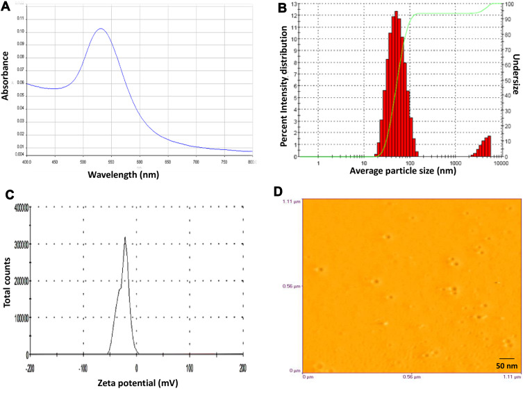
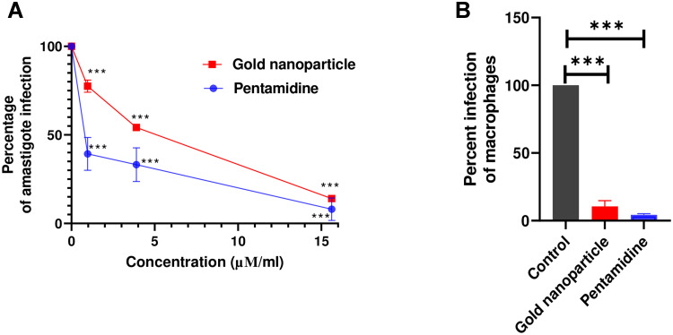
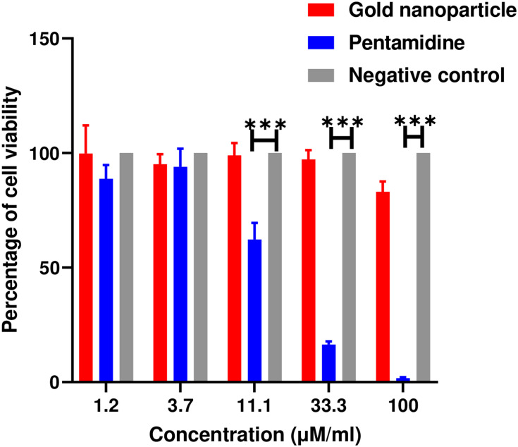
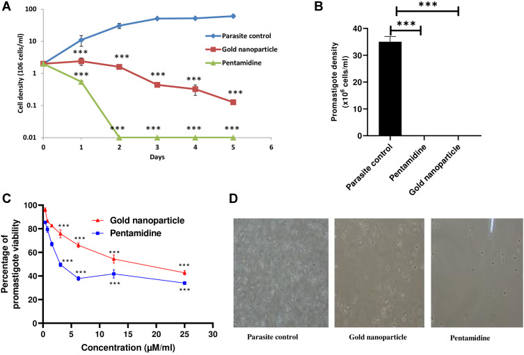
GNP showed a strong SPR peak at 520 nm and mean particle size, polydispersity index (PDI), and zeta potential of 56.0 ± 10 nm, 0.3 ± 0.1 and -27.0 ± 3 mV, respectively. The GNPs were smooth and spherical with a mean particle diameter of 20 ± 5 nm. Nanoparticles [1.2-100 µM] did not reveal any cytotoxicity on RAW 264.7 murine macrophage cell line, but exerted significant activity against both promastigotes and amastigote stages of with 50% inhibitory concentrations (IC) of 18.4 ± 0.4 µM and 5.0 ± 0.3 µM, respectively. GNP showed significant antileishmanial activity with deformed morphology of parasites and the least number of surviving promastigotes after growth reversibility analysis.
GNP may provide a platform to conjugate antileishmanial drugs onto the surface of nanoparticles to enhance their therapeutic effectiveness against VL. Further work is warranted, involving more in-depth mechanistic studies and in vivo investigations.
由 复合体引起的内脏利什曼病(VL)目前的治疗方法有限,并且存在严重的局限性。由于耐药性的增加,联合疗法已被证明无效;然而,在没有疫苗的情况下,寻找安全有效的药物是可取的。人们对纳米粒子在利什曼病治疗效果中的应用越来越感兴趣。鉴于此,我们评估了金纳米粒子(GNP)对 的体外抗利什曼效果。
通过动态光散射(DLS)和原子力显微镜(AFM)对 GNP 的粒径进行了合成和表征,并通过紫外-可见光谱对其光学性质进行了测量。通过 MTT 增殖测定法测量 GNP 的细胞毒性。评估了纳米粒子对体外 前体和巨噬细胞感染的无鞭毛体的抗利什曼活性。
GNP 在 520nm 处显示出很强的 SPR 峰,平均粒径、多分散指数(PDI)和 Zeta 电位分别为 56.0±10nm、0.3±0.1 和-27.0±3mV。GNPs 光滑呈球形,平均粒径为 20±5nm。纳米粒子[1.2-100µM]对 RAW 264.7 鼠巨噬细胞系没有显示出任何细胞毒性,但对前体和无鞭毛体阶段的 都具有显著的活性,半抑制浓度(IC)分别为 18.4±0.4µM 和 5.0±0.3µM。GNP 表现出显著的抗利什曼活性,表现为寄生虫形态变形,生长可逆性分析后存活的前体数量最少。
GNP 可为将抗利什曼药物共轭到纳米粒子表面提供平台,以增强其对 VL 的治疗效果。需要进一步的工作,包括更深入的机制研究和体内研究。