Department of General Practice, The First Affiliated Hospital of Hainan Medical University, Haikou, Hainan 570100, P.R. China.
Hainan Medical University, Haikou, Hainan 570100, P.R. China.
Mol Med Rep. 2022 Jan;25(1). doi: 10.3892/mmr.2021.12520. Epub 2021 Nov 5.
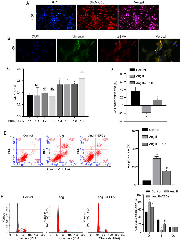
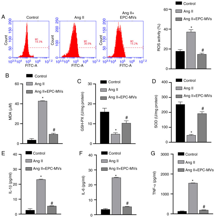
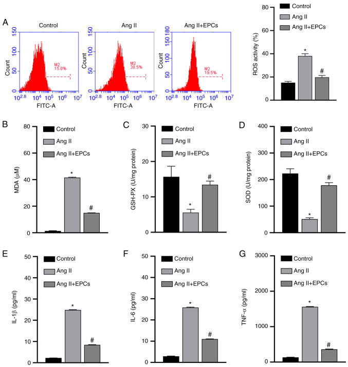
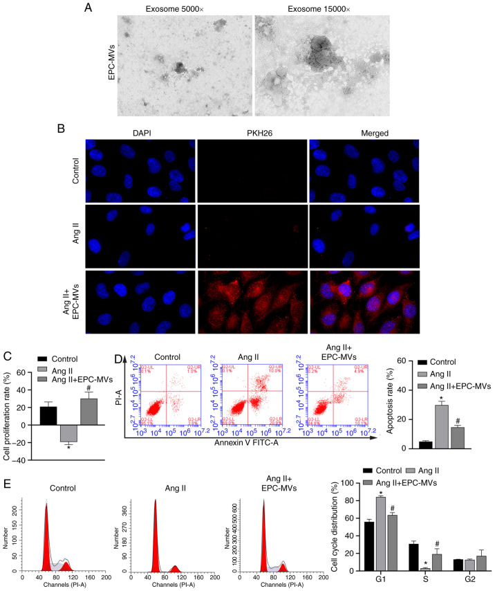
Chronic hypertension can lead to kidney damage, known as hypertensive nephropathy or hypertensive nephrosclerosis. Further understanding of the molecular mechanisms via which hypertensive nephropathy develops is essential for effective diagnosis and treatment. The present study investigated the mechanisms by which endothelial progenitor cells (EPCs) repair primary rat kidney cells (PRKs). ELISA, Cell Counting Kit‑8 and flow cytometry assays were used to analyze the effects of EPCs or EPC‑MVs on the oxidative stress, inflammation, cell proliferation, apoptosis and cycle of PRKs induced by AngII. A PRK injury model was established using angiotensin II (Ang II). After Ang II induction, PRK proliferation was decreased, apoptosis was increased and the cell cycle was blocked at the G phase before entering the S phase. It was found that the levels of reactive oxygen species and malondialdehyde were increased, while the levels of glutathione peroxidase and superoxide dismutase were decreased. Moreover, the levels of the inflammatory cytokines IL‑1β, IL‑6 and TNF‑α were significantly increased. Thus, Ang II damaged PRKs by stimulating oxidative stress and promoting the inflammatory response. However, when PRKs were co‑cultured with EPCs, the damage induced by Ang II was significantly reduced. The current study collected the microvesicles (MVs) secreted by EPCs and co‑cultured them with Ang II‑induced PRKs, and identified that EPC‑MVs retained their protective effect on PRKs. In conclusion, EPCs protect PRKs from Ang II‑induced damage via secreted MVs.
慢性高血压可导致肾脏损伤,即高血压肾病或高血压肾硬化症。进一步了解高血压肾病发展的分子机制对于有效诊断和治疗至关重要。本研究探讨了内皮祖细胞(EPC)修复原代大鼠肾细胞(PRK)的机制。ELISA、细胞计数试剂盒-8 和流式细胞术检测用于分析 EPC 或 EPC-MVs 对 AngII 诱导的 PRK 氧化应激、炎症、细胞增殖、凋亡和周期的影响。使用血管紧张素 II(Ang II)建立 PRK 损伤模型。Ang II 诱导后,PRK 增殖减少,凋亡增加,细胞周期在进入 S 期之前在 G 期停滞。结果发现,活性氧和丙二醛的水平升高,而谷胱甘肽过氧化物酶和超氧化物歧化酶的水平降低。此外,炎症细胞因子 IL-1β、IL-6 和 TNF-α 的水平显著升高。因此,Ang II 通过刺激氧化应激和促进炎症反应来损伤 PRK。然而,当 PRK 与 EPC 共培养时,Ang II 诱导的损伤明显减轻。本研究收集了 EPC 分泌的微泡(MVs)并与 Ang II 诱导的 PRK 共培养,结果表明 EPC-MVs 保留了对 PRK 的保护作用。总之,EPC 通过分泌的 MV 保护 PRK 免受 Ang II 诱导的损伤。