Department of Neurology, University of California Los Angeles, CA 90095, USA; Department of Biomedical Engineering, University of North Texas, TX 76207, USA.
Department of Neurology, University of California Los Angeles, CA 90095, USA.
Neurobiol Dis. 2021 Dec;161:105544. doi: 10.1016/j.nbd.2021.105544. Epub 2021 Nov 3.
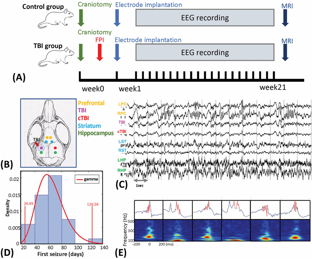
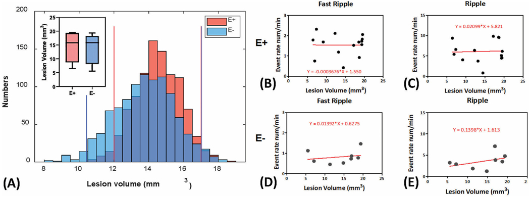
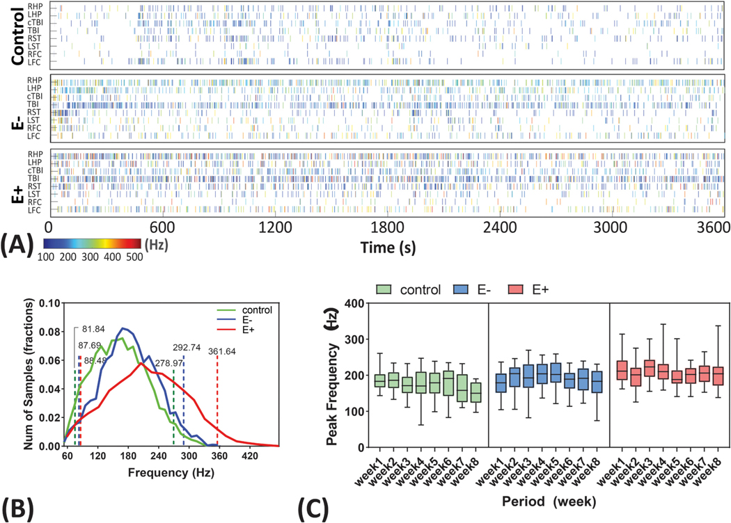
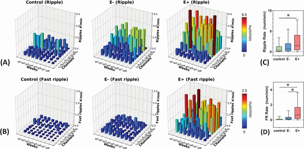
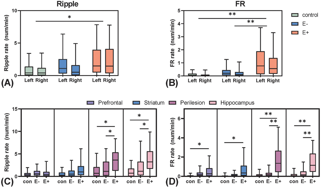
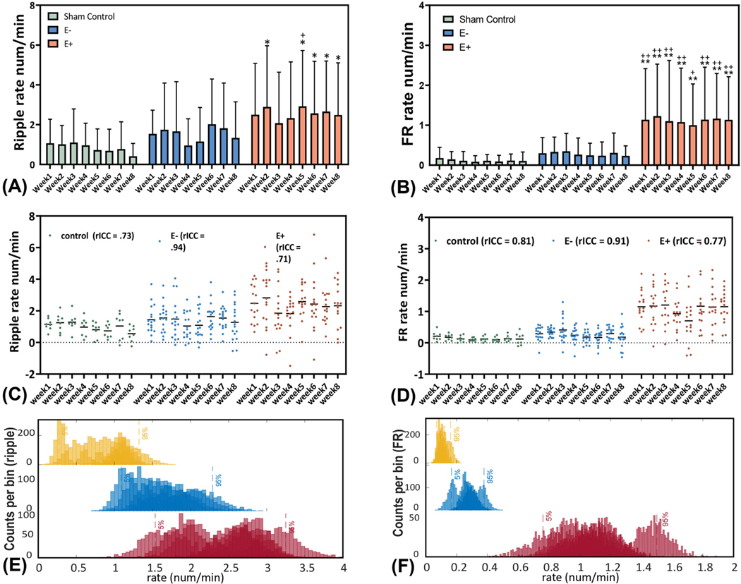
We studied the role of temporal and spatial changes in high-frequency oscillation (HFO, 80-500 Hz) generation in epileptogenesis following traumatic brain injury (TBI). Experiments were conducted on adult male Sprague Dawley rats. For the TBI group, fluid percussion injury (FPI) on the left sensorimotor area was performed to induce posttraumatic epileptogenesis. For the sham control group, only the craniotomy was performed. After TBI, 8 bipolar micro-electrodes were implanted bilaterally in the prefrontal cortex, perilesional area and homotopic contralateral site, striatum, and hippocampus. Long-term video/local field potential (LFP) recordings were performed for up to 21 weeks to identify and characterize seizures and capture HFOs. The electrode tip locations and the volume of post TBI brain lesions were further estimated by ex-vivo MRI scans. HFOs were detected during slow-wave sleep and categorized as ripple (80-200 Hz) and fast ripple (FR, 250-500 Hz) events. HFO rates and the HFO peak frequencies were compared in the 8 recording locations and across 8-weeks following TBI. Data from 48 rats (8 sham controls and 40 TBI rats) were analyzed. Within the TBI group, 22 rats (55%) developed recurrent spontaneous seizures (E+ group), at an average of 62.2 (+17.1) days, while 18 rats (45%) did not (E- group). We observed that the HFOs in the E+ group had a higher mean peak frequency than the E- group and the sham group (P < 0.05). Furthermore, the FR rate of the E+ group showed a significant increase compared to the E-group (P < 0.01) and sham control group (P < 0.01), specifically in the perilesional area, homotopic contralateral site, bilateral hippocampus, and to a lesser degree bilateral striatum. When compared across time, the increased FR rate in the E+ group occurred immediately after the insult and remained stable across the duration of the experiment. In addition, lesion size was not statistically different in the E+ and E- group and was not correlated with HFO rates. Our results suggest that TBI results in the formation of a widespread epileptogenic network. FR rates serve as a biomarker of network formation and predict the future development of epilepsy, however FR are not a temporally specific biomarker of TBI sequelae responsible for epileptogenesis. These results suggest that in patients, future risk of post-TBI epilepsy can be predicted early using FR.
我们研究了创伤性脑损伤(TBI)后癫痫发生时高频振荡(HFO,80-500 Hz)产生的时空变化的作用。实验在成年雄性 Sprague Dawley 大鼠上进行。对于 TBI 组,在左侧感觉运动区进行液压冲击伤(FPI)以诱导创伤后癫痫发生。对于假手术对照组,仅进行开颅术。TBI 后,在额皮质、损伤周围区域和对侧同部位、纹状体和海马双侧植入 8 个双极微电极。进行长达 21 周的长期视频/局部场电位(LFP)记录,以识别和特征化癫痫发作并捕获 HFO。通过离体 MRI 扫描进一步估计电极尖端位置和 TBI 后脑损伤的体积。在慢波睡眠期间检测到 HFO,并分类为锐波(80-200 Hz)和快锐波(FR,250-500 Hz)事件。比较了 TBI 后 8 周内 8 个记录位置的 HFO 率和 HFO 峰值频率。分析了 48 只大鼠(8 只假手术对照和 40 只 TBI 大鼠)的数据。在 TBI 组中,22 只大鼠(55%)出现复发性自发性癫痫发作(E+组),平均时间为 62.2(+17.1)天,而 18 只大鼠(45%)未出现(E-组)。我们观察到,E+组的 HFO 平均峰值频率高于 E-组和假手术组(P<0.05)。此外,E+组的 FR 率与 E-组(P<0.01)和假手术对照组(P<0.01)相比显著增加,特别是在损伤周围区域、对侧同部位、双侧海马体,并且在双侧纹状体的程度较小。与时间相比,E+组的 FR 率在损伤后立即增加,并在整个实验过程中保持稳定。此外,E+和 E-组的损伤大小没有统计学差异,并且与 HFO 率无关。我们的结果表明,TBI 导致形成广泛的致痫网络。FR 率可作为网络形成的生物标志物,并预测癫痫的未来发展,但是 FR 并不是导致癫痫发生的 TBI 后遗症的时间特异性生物标志物。这些结果表明,在患者中,可以使用 FR 早期预测 TBI 后癫痫的发生风险。