Sand Daniel, Arkadir David, Abu Snineh Muneer, Marmor Odeya, Israel Zvi, Bergman Hagai, Hassin-Baer Sharon, Israeli-Korn Simon, Peremen Ziv, Geva Amir B, Eitan Renana
Department of Medical Neurobiology (Physiology), Institute of Medical Research Israel-Canada, Hebrew University of Jerusalem, Jerusalem, Israel.
Edmond and Lily Safra Center for Brain Research, Hebrew University of Jerusalem, Jerusalem, Israel.
Front Syst Neurosci. 2021 Oct 20;15:747681. doi: 10.3389/fnsys.2021.747681. eCollection 2021.
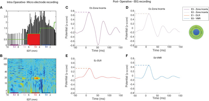
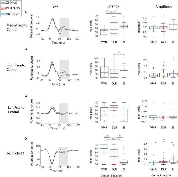
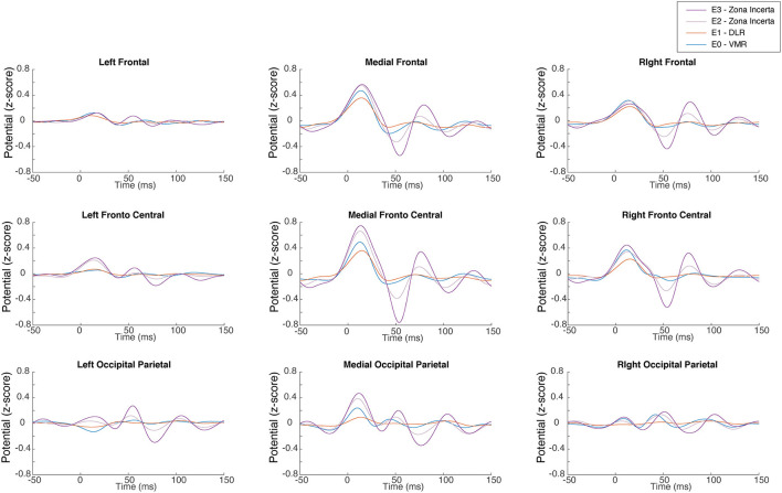
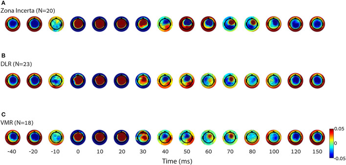
Precise lead localization is crucial for an optimal clinical outcome of subthalamic nucleus (STN) deep brain stimulation (DBS) treatment in patients with Parkinson's disease (PD). Currently, anatomical measures, as well as invasive intraoperative electrophysiological recordings, are used to locate DBS electrodes. The objective of this study was to find an alternative electrophysiology tool for STN DBS lead localization. Sixty-one postoperative electrophysiology recording sessions were obtained from 17 DBS-treated patients with PD. An intraoperative physiological method automatically detected STN borders and subregions. Postoperative EEG cortical activity was measured, while STN low frequency stimulation (LFS) was applied to different areas inside and outside the STN. Machine learning models were used to differentiate stimulation locations, based on EEG analysis of engineered features. A machine learning algorithm identified the top 25 evoked response potentials (ERPs), engineered features that can differentiate inside and outside STN stimulation locations as well as within STN stimulation locations. Evoked responses in the medial and ipsilateral fronto-central areas were found to be most significant for predicting the location of STN stimulation. Two-class linear support vector machine (SVM) predicted the inside (dorso-lateral region, DLR, and ventro-medial region, VMR) vs. outside [zona incerta, ZI, STN stimulation classification with an accuracy of 0.98 and 0.82 for ZI vs. VMR and ZI vs. DLR, respectively, and an accuracy of 0.77 for the within STN (DLR vs. VMR)]. Multiclass linear SVM predicted all areas with an accuracy of 0.82 for the outside and within STN stimulation locations (ZI vs. DLR vs. VMR). Electroencephalogram biomarkers can use low-frequency STN stimulation to localize STN DBS electrodes to ZI, DLR, and VMR STN subregions. These models can be used for both intraoperative electrode localization and postoperative stimulation programming sessions, and have a potential to improve STN DBS clinical outcomes.
精确的电极定位对于帕金森病(PD)患者丘脑底核(STN)深部脑刺激(DBS)治疗的最佳临床效果至关重要。目前,解剖学测量以及术中侵入性电生理记录被用于定位DBS电极。本研究的目的是找到一种用于STN DBS电极定位的替代电生理工具。从17例接受DBS治疗的PD患者中获得了61次术后电生理记录。一种术中生理方法自动检测STN边界和子区域。在对STN内部和外部的不同区域进行STN低频刺激(LFS)时,测量术后脑电图皮质活动。基于对工程特征的脑电图分析,使用机器学习模型来区分刺激位置。一种机器学习算法识别出前25个诱发反应电位(ERP),这些工程特征可以区分STN刺激位置的内部和外部以及STN刺激位置内的区域。发现内侧和同侧额中央区域的诱发反应对于预测STN刺激位置最为显著。两类线性支持向量机(SVM)预测内侧(背外侧区域,DLR,和腹内侧区域,VMR)与外侧[未定带,ZI,STN刺激分类,ZI与VMR以及ZI与DLR的准确率分别为0.98和0.82,STN内(DLR与VMR)的准确率为0.77]。多类线性SVM预测所有区域,STN刺激位置的内部和外部(ZI与DLR与VMR)的准确率为0.82。脑电图生物标志物可以利用低频STN刺激将STN DBS电极定位到ZI、DLR和VMR STN子区域。这些模型可用于术中电极定位和术后刺激编程,并且有潜力改善STN DBS的临床效果。