Department of Neurosurgery, Washington University in St. Louis School of Medicine, St. Louis, MO, 63110, USA.
Department of Neurosurgery, BJC Institute of Health, 425 S. Euclid, Campus, Box 8057, St. Louis, MO, 63143, USA.
Fluids Barriers CNS. 2021 Nov 8;18(1):49. doi: 10.1186/s12987-021-00281-0.
Many animal models have been used to study the pathophysiology of hydrocephalus; most of these have been rodent models whose lissencephalic cerebral cortex may not respond to ventriculomegaly in the same way as gyrencephalic species and whose size is not amenable to evaluation of clinically relevant neurosurgical treatments. Fewer models of hydrocephalus in gyrencephalic species have been used; thus, we have expanded upon a porcine model of hydrocephalus in juvenile pigs and used it to explore surgical treatment methods.
Acquired hydrocephalus was induced in 33-41-day old pigs by percutaneous intracisternal injections of kaolin (n = 17). Controls consisted of sham saline-injected (n = 6) and intact (n = 4) animals. Magnetic resonance imaging (MRI) was employed to evaluate ventriculomegaly at 11-42 days post-kaolin and to plan the surgical implantation of ventriculoperitoneal shunts at 14-38-days post-kaolin. Behavioral and neurological status were assessed.
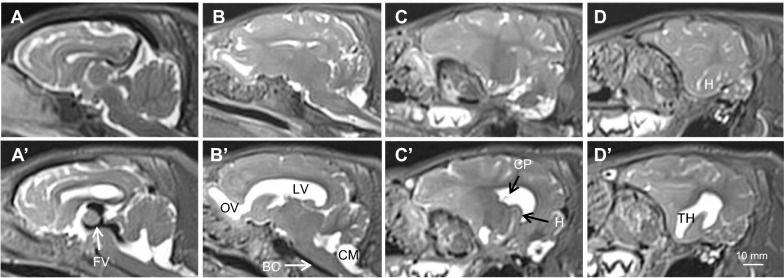
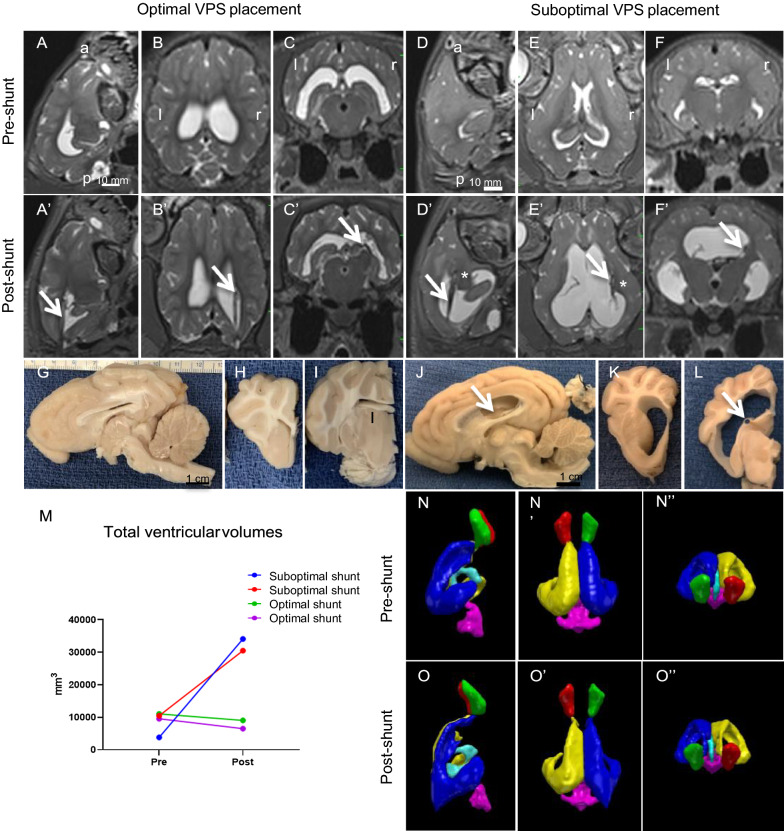
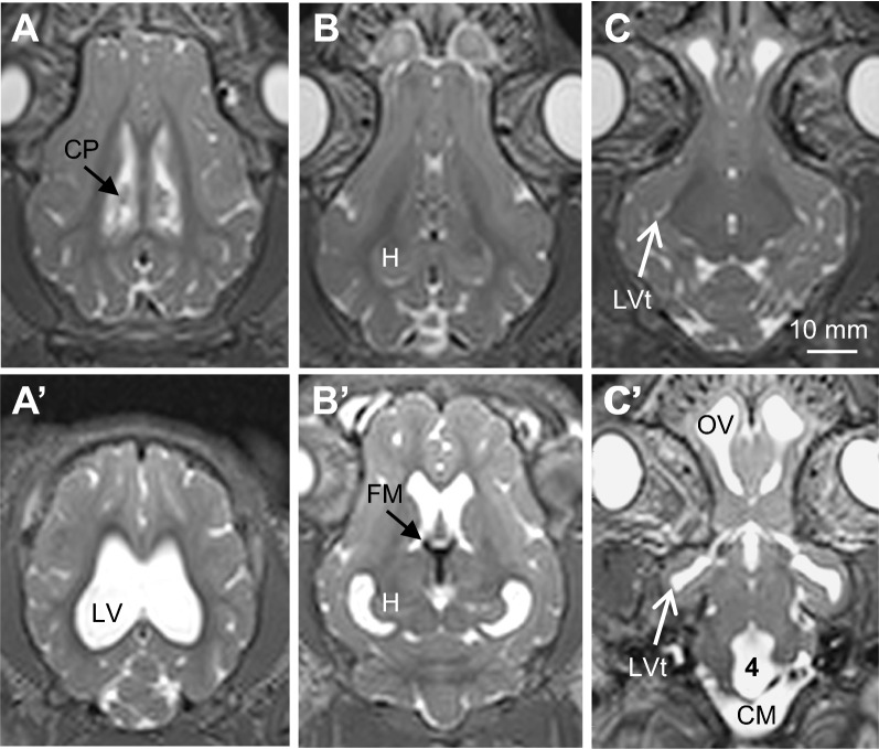
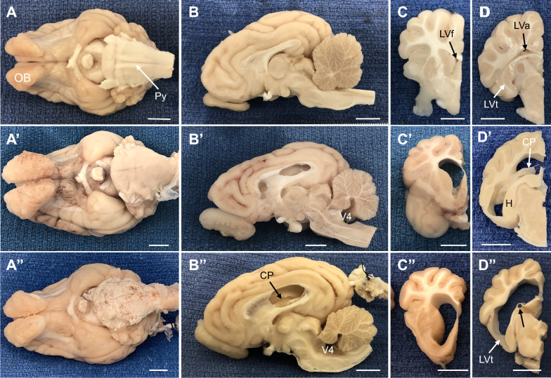
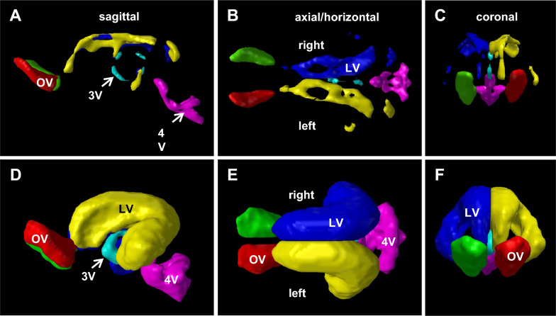
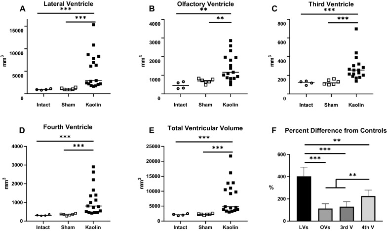
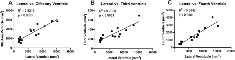
Bilateral ventriculomegaly occurred post-induction in all regions of the cerebral ventricles, with prominent CSF flow voids in the third ventricle, foramina of Monro, and cerebral aqueduct. Kaolin deposits formed a solid cast in the basal cisterns but the cisterna magna was patent. In 17 untreated hydrocephalic animals. Mean total ventricular volume was 8898 ± 5917 SD mm at 11-43 days of age, which was significantly larger than the baseline values of 2251 ± 194 SD mm for 6 sham controls aged 45-55 days, (p < 0.001). Past the post-induction recovery period, untreated pigs were asymptomatic despite exhibiting mild-moderate ventriculomegaly. Three out of 4 shunted animals showed a reduction in ventricular volume after 20-30 days of treatment, however some developed ataxia and lethargy, from putative shunt malfunction.
Kaolin induction of acquired hydrocephalus in juvenile pigs produced an in vivo model that is highly translational, allowing systematic studies of the pathophysiology and clinical treatment of hydrocephalus.
许多动物模型已被用于研究脑积水的病理生理学;其中大多数是啮齿动物模型,其脑回较少的大脑皮质可能不会像脑回较多的物种那样对脑室扩大产生相同的反应,而且其大小也不适合评估临床相关的神经外科治疗。脑积水的脑回较多的物种模型使用较少;因此,我们在幼年猪中扩展了脑积水的猪模型,并使用它来探索手术治疗方法。
通过经皮脑室内注射高岭土在 33-41 天大的猪中诱发获得性脑积水(n=17)。对照组包括假盐水注射(n=6)和完整(n=4)动物。磁共振成像(MRI)用于评估高岭土后 11-42 天的脑室扩大,并计划在高岭土后 14-38 天进行脑室-腹腔分流术的手术植入。评估行为和神经状态。
诱导后所有脑室内区域均出现双侧脑室扩大,第三脑室、Monro 孔和脑导水管出现明显的 CSF 流空。高岭土沉积物在基底池内形成实心铸型,但大脑大静脉池保持通畅。在 17 只未经治疗的脑积水动物中,平均总脑室容积在 11-43 天大时为 8898±5917 SD mm,明显大于 6 只 45-55 天大的假对照的基线值 2251±194 SD mm(p<0.001)。在诱导后恢复期间,尽管存在轻度至中度脑室扩大,但未经治疗的猪无症状。4 只分流动物中有 3 只在 20-30 天治疗后脑室容积减少,但有些出现共济失调和昏睡,可能是分流器故障所致。
高岭土诱导幼年猪获得性脑积水产生了一种高度转化的体内模型,允许对脑积水的病理生理学和临床治疗进行系统研究。