Zaitun Hasibuan Poppy Anjelisa, Tanjung Masitta, Gea Saharman, Pasaribu Khatarina Meldawati, Harahap Mahyuni, Perangin-Angin Yurika Almanda, Prayoga Andre, Ginting Junius Gian
Department of Pharmacology, Faculty of Pharmacy, Universitas Sumatera Utara, Jl. Tridharma No.5, Medan, 22919, Indonesia.
Department of Biology, Faculty of Mathematics and Natural Science, Universitas Sumatera Utara, Jl. Bioteknologi No.1, Medan, 20155, Indonesia.
Heliyon. 2021 Oct 19;7(10):e08197. doi: 10.1016/j.heliyon.2021.e08197. eCollection 2021 Oct.
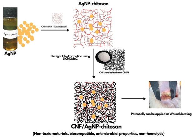
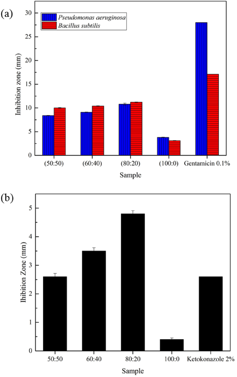
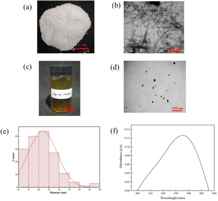
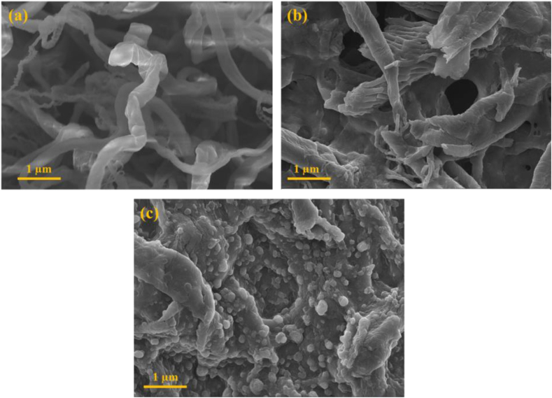
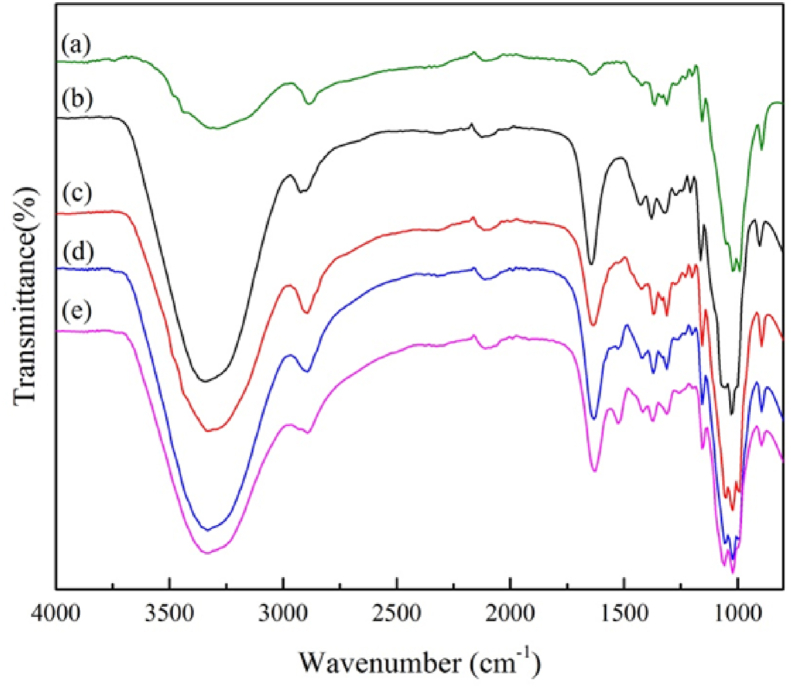
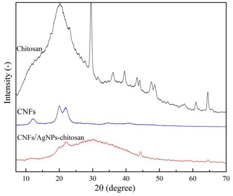
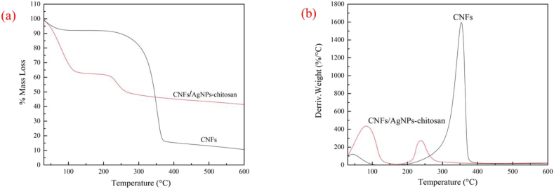
Cellulose nanofibers (CNFs), chitosan, and silver nanoparticles (AgNPs) are widely used to enhance the active functions and antibacterial properties of wound dressings. This study was conducted to prepare CNF/AgNP-chitosan using a straight incorporation method and to assess its antimicrobial activity. CNFs were isolated from oil palm empty fruit bunches (OPEFBs) using the pulping method and acid hydrolysis. AgNPs were synthesized using a green synthesis method. The wound dressing was produced by mixing a 10% CNF solution in LiCl/DMAc and AgNP-chitosan in a glass plate with various ratios of CNF/AgNP-chitosan, i.e., 100:0, 80:20, 60:40, and 50:50. UV-visible and TEM analyses were conducted to confirm the formation of AgNPs and CNFs at the nanoscale. The results showed particles with an absorption wavelength of 435 nm and spherical shapes. Based on the calculation using ImageJ software, the diameters of CNFs were approximately 50 nm, and the lengths were several micrometers. FTIR was used to analyze the chemical bonding of AgNP-chitosan and the incorporated AgNP-chitosan in CNFs. Based on the XRD analysis, the presence of AgNPs did not affect the crystallinity of the CNFs. SEM images showed that the addition of AgNPs resulted in the stretching of CNF pores on the pads. Thermal degradation of the film increased with the addition of AgNP-chitosan by up to 40%. Antimicrobial tests and hemocompatibility tests showed that the formed CNF/AgNP-chitosan film successfully inhibited bacterial growth and was classified as a nonhemolytic material; thus, its potential as a wound dressing should be further studied.
纤维素纳米纤维(CNFs)、壳聚糖和银纳米颗粒(AgNPs)被广泛用于增强伤口敷料的活性功能和抗菌性能。本研究采用直接掺入法制备了CNF/AgNP-壳聚糖,并评估其抗菌活性。通过制浆法和酸水解从油棕空果串(OPEFBs)中分离出CNFs。采用绿色合成法合成AgNPs。通过将10%的CNF溶液在LiCl/DMAc中与AgNP-壳聚糖以不同比例(即100:0、80:20、60:40和50:50)在玻璃板上混合来制备伤口敷料。进行紫外可见和透射电镜分析以确认纳米级AgNPs和CNFs的形成。结果显示颗粒的吸收波长为435 nm且呈球形。基于使用ImageJ软件的计算,CNFs的直径约为50 nm,长度为几微米。傅里叶变换红外光谱(FTIR)用于分析AgNP-壳聚糖的化学键以及掺入CNFs中的AgNP-壳聚糖。基于X射线衍射(XRD)分析,AgNPs的存在不影响CNFs的结晶度。扫描电子显微镜(SEM)图像显示添加AgNPs导致垫上CNF孔隙的拉伸。随着AgNP-壳聚糖的添加,薄膜的热降解增加了高达40%。抗菌测试和血液相容性测试表明,形成的CNF/AgNP-壳聚糖薄膜成功抑制了细菌生长,并且被归类为非溶血材料;因此,其作为伤口敷料的潜力应进一步研究。