• 文献检索
  • 文档翻译
  • 深度研究
  • 学术资讯
  • Suppr Zotero 插件Zotero 插件
  • 邀请有礼
  • 套餐&价格
  • 历史记录
应用&插件
Suppr Zotero 插件Zotero 插件浏览器插件Mac 客户端Windows 客户端微信小程序
定价
高级版会员购买积分包购买API积分包
服务
文献检索文档翻译深度研究API 文档MCP 服务
关于我们
关于 Suppr公司介绍联系我们用户协议隐私条款
关注我们

Suppr 超能文献

核心技术专利:CN118964589B侵权必究
粤ICP备2023148730 号-1Suppr @ 2026

文献检索

告别复杂PubMed语法,用中文像聊天一样搜索,搜遍4000万医学文献。AI智能推荐,让科研检索更轻松。

立即免费搜索

文件翻译

保留排版,准确专业,支持PDF/Word/PPT等文件格式,支持 12+语言互译。

免费翻译文档

深度研究

AI帮你快速写综述,25分钟生成高质量综述,智能提取关键信息,辅助科研写作。

立即免费体验

入院时计算机断层扫描检查发现的脂肪肝是严重冠状病毒疾病的危险因素。

Fatty liver on computed tomography scan on admission is a risk factor for severe coronavirus disease.

机构信息

Disease Control and Prevention Center, National Center for Global Health and Medicine, Tokyo, Japan.

Department of Radiology, National Center for Global Health and Medicine, Tokyo, Japan.

出版信息

J Infect Chemother. 2022 Feb;28(2):217-223. doi: 10.1016/j.jiac.2021.10.013. Epub 2021 Oct 20.

DOI:10.1016/j.jiac.2021.10.013
PMID:34756574
原文链接:https://pmc.ncbi.nlm.nih.gov/articles/PMC8526433/
Abstract

OBJECTIVES

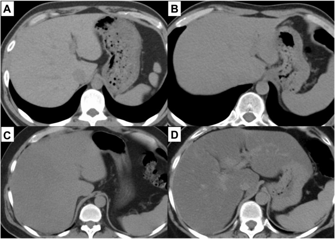
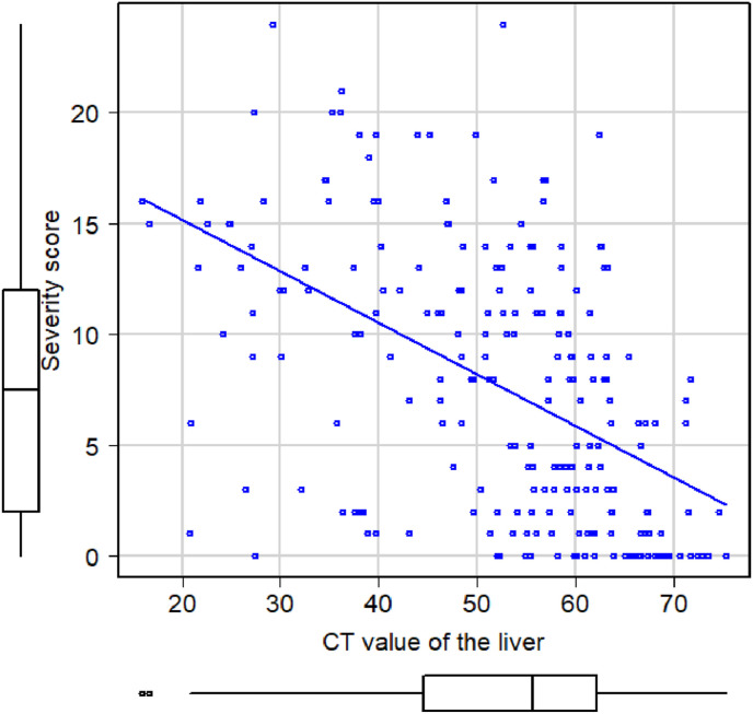
To alleviate the overflow of coronavirus disease 2019 (COVID-19) patients in hospitals, less invasive and simple criteria are required to triage the patients. We evaluated the relationship between COVID-19 severity and fatty liver on plain computed tomography (CT) scan performed on admission.

METHODS

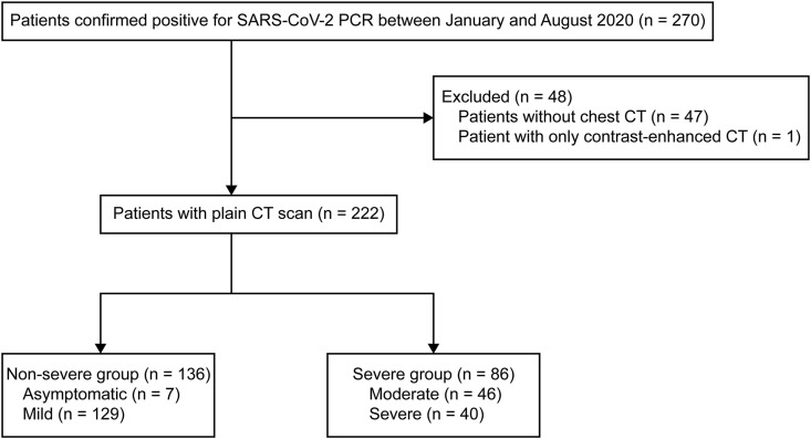
In this retrospective cohort study, we considered all COVID-19 patients at a large tertiary care hospital between January 31 and August 31, 2020. COVID-19 severity was categorized into severe (moderate and severe) and non-severe (asymptomatic and mild) groups, based on the Japanese National COVID-19 guidelines. Fatty liver was detected on plain CT scan. Multivariate logistic regression analysis was performed to evaluate factors associated with severe COVID-19.

RESULTS

Of 222 patients (median age: 52 years), 3.2%, 58.1%, 20.7%, and 18.0% presented with asymptomatic, mild, moderate, and severe COVID-19, respectively. Although 59.9% had no fatty liver on plain CT, mild, moderate, and severe fatty liver occurred in 13.1%, 18.9%, and 8.1%, respectively. Age and presence of fatty liver were significantly associated with severe COVID-19.

CONCLUSION

Our study showed that fatty liver on plain CT scan on admission can become a risk factor for severe COVID-19. This finding may help clinicians to easily triage COVID-19 patients.

摘要

目的

为缓解医院 2019 冠状病毒病(COVID-19)患者的积压,需要采用侵入性更小、更简单的标准对患者进行分诊。我们评估了入院时普通计算机断层扫描(CT)上的脂肪肝与 COVID-19 严重程度之间的关系。

方法

在这项回顾性队列研究中,我们考虑了 2020 年 1 月 31 日至 8 月 31 日期间在一家大型三级保健医院的所有 COVID-19 患者。根据日本国家 COVID-19 指南,将 COVID-19 严重程度分为严重(中度和重度)和非严重(无症状和轻度)两组。在普通 CT 扫描上检测脂肪肝。采用多变量逻辑回归分析评估与严重 COVID-19 相关的因素。

结果

在 222 例患者中(中位年龄:52 岁),分别有 3.2%、58.1%、20.7%和 18.0%的患者为无症状、轻度、中度和重度 COVID-19。尽管 59.9%的患者在普通 CT 上没有脂肪肝,但分别有 13.1%、18.9%和 8.1%的患者有轻度、中度和重度脂肪肝。年龄和脂肪肝的存在与严重 COVID-19 显著相关。

结论

我们的研究表明,入院时普通 CT 扫描上的脂肪肝可能成为严重 COVID-19 的危险因素。这一发现可能有助于临床医生对 COVID-19 患者进行简单分诊。

https://cdn.ncbi.nlm.nih.gov/pmc/blobs/7d4f/8526433/426b1d949bdb/gr3_lrg.jpg
https://cdn.ncbi.nlm.nih.gov/pmc/blobs/7d4f/8526433/73cb0b293eb8/gr1_lrg.jpg
https://cdn.ncbi.nlm.nih.gov/pmc/blobs/7d4f/8526433/fab8c23e6353/gr2_lrg.jpg
https://cdn.ncbi.nlm.nih.gov/pmc/blobs/7d4f/8526433/426b1d949bdb/gr3_lrg.jpg
https://cdn.ncbi.nlm.nih.gov/pmc/blobs/7d4f/8526433/73cb0b293eb8/gr1_lrg.jpg
https://cdn.ncbi.nlm.nih.gov/pmc/blobs/7d4f/8526433/fab8c23e6353/gr2_lrg.jpg
https://cdn.ncbi.nlm.nih.gov/pmc/blobs/7d4f/8526433/426b1d949bdb/gr3_lrg.jpg

相似文献

1
Fatty liver on computed tomography scan on admission is a risk factor for severe coronavirus disease.入院时计算机断层扫描检查发现的脂肪肝是严重冠状病毒疾病的危险因素。
J Infect Chemother. 2022 Feb;28(2):217-223. doi: 10.1016/j.jiac.2021.10.013. Epub 2021 Oct 20.
2
CT features of SARS-CoV-2 pneumonia according to clinical presentation: a retrospective analysis of 120 consecutive patients from Wuhan city.根据临床表现的 SARS-CoV-2 肺炎的 CT 特征:对武汉市 120 例连续患者的回顾性分析。
Eur Radiol. 2020 Aug;30(8):4417-4426. doi: 10.1007/s00330-020-06854-1. Epub 2020 Apr 11.
3
The effect of hepatic steatosis on COVID-19 severity: Chest computed tomography findings.肝脂肪变性对 COVID-19 严重程度的影响:胸部 CT 表现。
Saudi J Gastroenterol. 2021 Mar-Apr;27(2):105-110. doi: 10.4103/sjg.sjg_540_20.
4
Association and Interaction Between Serum Interleukin-6 Levels and Metabolic Dysfunction-Associated Fatty Liver Disease in Patients With Severe Coronavirus Disease 2019.血清白细胞介素-6 水平与 2019 年严重冠状病毒病患者代谢功能障碍相关脂肪性肝病的相关性和相互作用。
Front Endocrinol (Lausanne). 2021 Mar 8;12:604100. doi: 10.3389/fendo.2021.604100. eCollection 2021.
5
[Spatial and temporal distribution and predictive value of chest CT scoring in patients with COVID-19].新型冠状病毒肺炎患者胸部CT评分的时空分布及预测价值
Zhonghua Jie He He Hu Xi Za Zhi. 2021 Mar 12;44(3):230-236. doi: 10.3760/cma.j.cn112147-20200522-00626.
6
CT Scan Findings of Five Mildly Symptomatic COVID Patients from India.来自印度的5例轻症新冠患者的CT扫描结果
J Coll Physicians Surg Pak. 2020 Jun;30(6):48-49. doi: 10.29271/jcpsp.2020.Supp1.S48.
7
Dandelion and focal crazy paving signs: the lung CT based predictors for evaluation of the severity of coronavirus disease.蒲公英征和局灶性铺路石征:基于肺部 CT 的预测因子用于评估冠状病毒病的严重程度。
Curr Med Res Opin. 2021 Feb;37(2):219-224. doi: 10.1080/03007995.2020.1846173. Epub 2020 Nov 26.
8
Risk factors for adverse clinical outcomes with COVID-19 in China: a multicenter, retrospective, observational study.中国 COVID-19 不良临床结局的危险因素:一项多中心、回顾性、观察性研究。
Theranostics. 2020 May 15;10(14):6372-6383. doi: 10.7150/thno.46833. eCollection 2020.
9
Understanding factors influencing the length of hospital stay among non-severe COVID-19 patients: A retrospective cohort study in a Fangcang shelter hospital.了解非重症 COVID-19 患者住院时间长短的影响因素:方仓庇护医院的回顾性队列研究。
PLoS One. 2020 Oct 21;15(10):e0240959. doi: 10.1371/journal.pone.0240959. eCollection 2020.
10
Comparative study of lung ultrasound and chest computed tomography scan in the assessment of severity of confirmed COVID-19 pneumonia.肺超声与胸部 CT 扫描在评估确诊 COVID-19 肺炎严重程度中的对比研究。
Intensive Care Med. 2020 Sep;46(9):1707-1713. doi: 10.1007/s00134-020-06186-0. Epub 2020 Jul 29.

引用本文的文献

1
Comparison of liver fibrosis scores and fatty liver on computed tomography as risk factors for severity of COVID-19.计算机断层扫描中肝纤维化评分和脂肪肝作为COVID-19严重程度风险因素的比较。
JGH Open. 2024 Aug 8;8(8):e70004. doi: 10.1002/jgh3.70004. eCollection 2024 Aug.
2
Non-alcoholic fatty liver disease (NAFLD) and COVID-19 outcomes: A systematic review, meta-analysis, and meta-regression.非酒精性脂肪性肝病(NAFLD)与2019冠状病毒病的转归:一项系统评价、荟萃分析和荟萃回归分析
Narra J. 2023 Apr;3(1):e102. doi: 10.52225/narra.v3i1.102. Epub 2023 Apr 19.
3
The Intersection of COVID-19 and Metabolic-Associated Fatty Liver Disease: An Overview of the Current Evidence.
新型冠状病毒肺炎与代谢相关脂肪性肝病的关系:当前证据概述。
Viruses. 2023 Apr 27;15(5):1072. doi: 10.3390/v15051072.
4
Non-alcoholic fatty liver disease and COVID-19: Harmless companions or disease intensifier?非酒精性脂肪性肝病与 COVID-19:无害伴侣还是疾病加剧因素?
World J Gastroenterol. 2023 Jan 14;29(2):367-377. doi: 10.3748/wjg.v29.i2.367.