• 文献检索
  • 文档翻译
  • 深度研究
  • 学术资讯
  • Suppr Zotero 插件Zotero 插件
  • 邀请有礼
  • 套餐&价格
  • 历史记录
应用&插件
Suppr Zotero 插件Zotero 插件浏览器插件Mac 客户端Windows 客户端微信小程序
定价
高级版会员购买积分包购买API积分包
服务
文献检索文档翻译深度研究API 文档MCP 服务
关于我们
关于 Suppr公司介绍联系我们用户协议隐私条款
关注我们

Suppr 超能文献

核心技术专利:CN118964589B侵权必究
粤ICP备2023148730 号-1Suppr @ 2026

文献检索

告别复杂PubMed语法,用中文像聊天一样搜索,搜遍4000万医学文献。AI智能推荐,让科研检索更轻松。

立即免费搜索

文件翻译

保留排版,准确专业,支持PDF/Word/PPT等文件格式,支持 12+语言互译。

免费翻译文档

深度研究

AI帮你快速写综述,25分钟生成高质量综述,智能提取关键信息,辅助科研写作。

立即免费体验

肝脂肪变性对 COVID-19 严重程度的影响:胸部 CT 表现。

The effect of hepatic steatosis on COVID-19 severity: Chest computed tomography findings.

机构信息

Department of Radiology, Ankara City Hospital, Turkey.

Department of Emergency Medicine, Ankara City Hospital, Turkey.

出版信息

Saudi J Gastroenterol. 2021 Mar-Apr;27(2):105-110. doi: 10.4103/sjg.sjg_540_20.

DOI:10.4103/sjg.sjg_540_20
PMID:33642355
原文链接:https://pmc.ncbi.nlm.nih.gov/articles/PMC8183358/
Abstract

BACKGROUND

The objective of our study was to investigate the location, extension and type of novel coronavirus-induced disease 2019 (COVID-19) infection involvement and hepatic steatosis on initial chest computed tomography (CT). The relationship between fatty liver and severity of the disease was also investigated by measuring the liver attenuation index (LAI).

METHODS

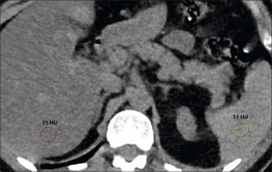
This study evaluated the chest CT images of 343 patients (201 male, mean age 48.43 years) who were confirmed to have COVID-19, using nasopharyngeal swab. The chest CTs were analyzed for laterality, number of involved lobes, diffuseness, number of lesions, and lesion types. The CT attenuation values of liver and spleen were measured, and LAI was calculated for the detection of hepatic steatosis. Univariate and multivariate logistic regression analysis were used to identify the independent early predictors for severe COVID-19.

RESULTS

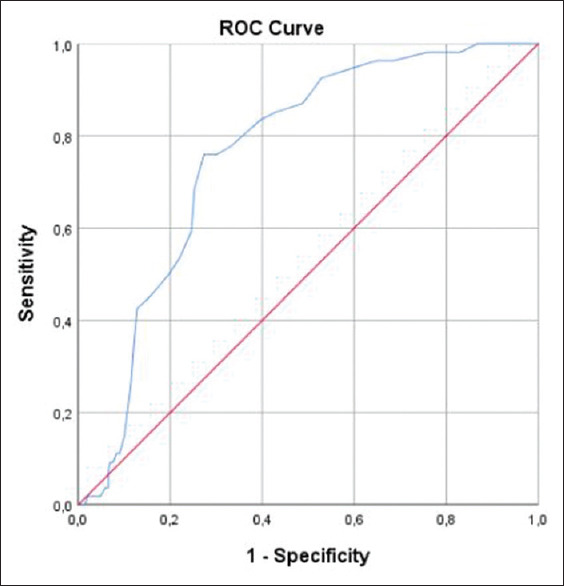
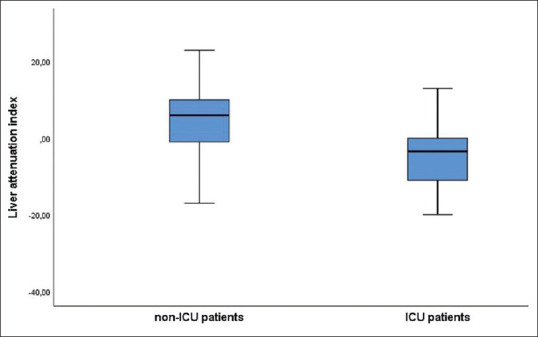
There was no significant difference between genders in terms of clinical course. Liver density and LAI were significantly lower in the intensive care unit (ICU) patients. The prevalence of severe disease was higher in the patients with hepatic steatosis than in the non-steatotic group (odds ratio [OR] 3.815, 95% confidence interval [CI] 1.97-7.37, P < 0.001). After adjusting for age and comorbidities including hypertension, diabetes mellitus, coronary artery disease, chronic obstructive pulmonary disease, and chronic kidney disease, multivariate logistic regression analysis showed that non-alcoholic fatty liver disease (NAFLD) was an independent risk factor for COVID-19 severity (OR 3.935, 95% CI 1.77-8.70, P = 0.001). The optimal cut-off value for LAI was calculated as 0.5 for predicting patients who required ICU treatment.

CONCLUSIONS

On the initial chest CT images of COVID-19 patients, presence of fatty liver is a strong predictor for severe disease.

摘要

背景

本研究旨在探讨 2019 年新型冠状病毒(COVID-19)感染引起的疾病在初始胸部计算机断层扫描(CT)中的位置、范围和类型,以及肝脂肪变性与疾病严重程度的关系,并通过测量肝脏衰减指数(LAI)来评估。

方法

本研究共评估了 343 例经鼻咽拭子确诊为 COVID-19 的患者的胸部 CT 图像(男 201 例,平均年龄 48.43 岁)。分析了病变的侧别、受累肺叶数、弥漫性、病变数和病变类型。测量了肝脏和脾脏的 CT 衰减值,并计算了 LAI 以检测肝脂肪变性。采用单因素和多因素逻辑回归分析确定严重 COVID-19 的独立早期预测因素。

结果

在临床病程方面,男女之间无显著性差异。在重症监护病房(ICU)患者中,肝脏密度和 LAI 显著降低。与非脂肪变性组相比,肝脂肪变性患者发生严重疾病的比例更高(比值比[OR] 3.815,95%置信区间[CI] 1.97-7.37,P <0.001)。在调整年龄和高血压、糖尿病、冠状动脉疾病、慢性阻塞性肺疾病和慢性肾脏病等合并症后,多因素逻辑回归分析显示,非酒精性脂肪性肝病(NAFLD)是 COVID-19 严重程度的独立危险因素(OR 3.935,95%CI 1.77-8.70,P = 0.001)。LAI 的最佳截断值为 0.5,可预测需要 ICU 治疗的患者。

结论

在 COVID-19 患者的初始胸部 CT 图像上,存在脂肪肝是疾病严重程度的一个强烈预测因素。

https://cdn.ncbi.nlm.nih.gov/pmc/blobs/676c/8183358/b7a509392c35/SJG-27-105-g003.jpg
https://cdn.ncbi.nlm.nih.gov/pmc/blobs/676c/8183358/31048f3269ca/SJG-27-105-g001.jpg
https://cdn.ncbi.nlm.nih.gov/pmc/blobs/676c/8183358/a2d6e6f3eaea/SJG-27-105-g002.jpg
https://cdn.ncbi.nlm.nih.gov/pmc/blobs/676c/8183358/b7a509392c35/SJG-27-105-g003.jpg
https://cdn.ncbi.nlm.nih.gov/pmc/blobs/676c/8183358/31048f3269ca/SJG-27-105-g001.jpg
https://cdn.ncbi.nlm.nih.gov/pmc/blobs/676c/8183358/a2d6e6f3eaea/SJG-27-105-g002.jpg
https://cdn.ncbi.nlm.nih.gov/pmc/blobs/676c/8183358/b7a509392c35/SJG-27-105-g003.jpg

相似文献

1
The effect of hepatic steatosis on COVID-19 severity: Chest computed tomography findings.肝脂肪变性对 COVID-19 严重程度的影响:胸部 CT 表现。
Saudi J Gastroenterol. 2021 Mar-Apr;27(2):105-110. doi: 10.4103/sjg.sjg_540_20.
2
Relationship of hepatic steatosis severity and coronary artery disease characteristics assessed by coronary CT angiography.通过冠状动脉CT血管造影评估肝脂肪变性严重程度与冠状动脉疾病特征的关系。
Int J Cardiovasc Imaging. 2016 Jun;32 Suppl 1:73-82. doi: 10.1007/s10554-016-0847-7. Epub 2016 Jan 30.
3
Frequency of hepatic steatosis and its association with the pneumonia severity score on chest computed tomography in adult COVID-19 patients.成年新冠病毒肺炎患者肝脏脂肪变性的发生率及其与胸部计算机断层扫描肺炎严重程度评分的相关性
World J Crit Care Med. 2021 May 9;10(3):47-57. doi: 10.5492/wjccm.v10.i3.47.
4
Higher frequency of hepatic steatosis at CT among COVID-19-positive patients.COVID-19 阳性患者 CT 检查中肝脂肪变性的频率更高。
Abdom Radiol (NY). 2020 Sep;45(9):2748-2754. doi: 10.1007/s00261-020-02648-7. Epub 2020 Jul 18.
5
Body Mass Index and Unenhanced CT as a Predictor of Hepatic Steatosis in Potential Liver Donors.体重指数和非增强CT作为潜在肝脏供体肝脂肪变性的预测指标
Transplant Proc. 2019 Sep;51(7):2373-2378. doi: 10.1016/j.transproceed.2019.02.047. Epub 2019 Aug 8.
6
Chest CT opportunistic biomarkers for phenotyping high-risk COVID-19 patients: a retrospective multicentre study.胸部 CT 机会性生物标志物用于表型分析 COVID-19 高危患者:一项回顾性多中心研究。
Eur Radiol. 2023 Nov;33(11):7756-7768. doi: 10.1007/s00330-023-09702-0. Epub 2023 May 11.
7
May an incidental finding on chest CT be a predictor of access in intensive care unit? Role of hepatic steatosis in patients affected by SARS-CoV-2.胸部CT的偶然发现能否作为重症监护病房入住的预测指标?肝脂肪变性在感染新型冠状病毒肺炎患者中的作用。
J Public Health Res. 2022 Nov 15;11(4):22799036221115779. doi: 10.1177/22799036221115779. eCollection 2022 Oct.
8
Prevalence of Hepatic Steatosis in Adults Presenting to the Emergency Department Identified by Unenhanced Chest CT.急诊就诊成年人中通过未增强胸部 CT 确定的肝脂肪变性患病率。
Curr Probl Diagn Radiol. 2023 Jan-Feb;52(1):35-40. doi: 10.1067/j.cpradiol.2022.07.014. Epub 2022 Jul 30.
9
Deep-learning-based hepatic fat assessment (DeHFt) on non-contrast chest CT and its association with disease severity in COVID-19 infections: A multi-site retrospective study.基于深度学习的非对比胸部 CT 肝脂肪评估(DeHFt)及其与 COVID-19 感染严重程度的关系:一项多中心回顾性研究。
EBioMedicine. 2022 Nov;85:104315. doi: 10.1016/j.ebiom.2022.104315. Epub 2022 Oct 26.
10
Virtual noncontrast images derived from dual-energy CT for assessment of hepatic steatosis in living liver donors.基于双能 CT 的虚拟平扫图像在活体肝移植供者肝脂肪变评估中的应用。
Eur J Radiol. 2021 Jun;139:109687. doi: 10.1016/j.ejrad.2021.109687. Epub 2021 Mar 27.

引用本文的文献

1
Comparison of liver fibrosis scores and fatty liver on computed tomography as risk factors for severity of COVID-19.计算机断层扫描中肝纤维化评分和脂肪肝作为COVID-19严重程度风险因素的比较。
JGH Open. 2024 Aug 8;8(8):e70004. doi: 10.1002/jgh3.70004. eCollection 2024 Aug.
2
Deep-learning segmentation to select liver parenchyma for categorizing hepatic steatosis on multinational chest CT.深度学习分割法选择肝脏实质用于对多国胸部 CT 上的肝脂肪变性进行分类。
Sci Rep. 2024 May 25;14(1):11987. doi: 10.1038/s41598-024-62887-2.
3
Is Steatotic Liver Disease Related to Poor Outcome in COVID-19-Hospitalized Patients?

本文引用的文献

1
COVID-19 and non-alcoholic fatty liver disease: Two intersecting pandemics.COVID-19 与非酒精性脂肪性肝病:两大流行病的交集。
Eur J Clin Invest. 2020 Oct;50(10):e13338. doi: 10.1111/eci.13338. Epub 2020 Aug 26.
2
Metabolic-associated fatty liver disease is associated with severity of COVID-19.代谢相关脂肪性肝病与 COVID-19 的严重程度相关。
Liver Int. 2020 Sep;40(9):2160-2163. doi: 10.1111/liv.14575. Epub 2020 Jul 5.
3
Review article: COVID-19 and liver disease-what we know on 1st May 2020.综述文章:2020 年 5 月 1 日的 COVID-19 和肝脏疾病——我们所知道的。
脂肪性肝病与COVID-19住院患者的不良预后有关吗?
J Clin Med. 2024 May 3;13(9):2687. doi: 10.3390/jcm13092687.
4
The Intersection of COVID-19 and Metabolic-Associated Fatty Liver Disease: An Overview of the Current Evidence.新型冠状病毒肺炎与代谢相关脂肪性肝病的关系:当前证据概述。
Viruses. 2023 Apr 27;15(5):1072. doi: 10.3390/v15051072.
5
Relationship between hepatic and pancreatic steatosis and the COVID-19 pneumonia total severity score and prognosis with an emphasis on prognostic strength.肝和胰腺脂肪变性与 COVID-19 肺炎总严重程度评分和预后的关系,重点是预后强度。
Diagn Interv Radiol. 2023 Jul 20;29(4):363-370. doi: 10.4274/dir.2022.221730. Epub 2022 Dec 21.
6
Non-alcoholic fatty liver disease and COVID-19: Harmless companions or disease intensifier?非酒精性脂肪性肝病与 COVID-19:无害伴侣还是疾病加剧因素?
World J Gastroenterol. 2023 Jan 14;29(2):367-377. doi: 10.3748/wjg.v29.i2.367.
7
Pancreatic Steatosis Evaluated by Automated Volumetric CT Fat Fraction of the Pancreas: Association with Severity in COVID-19 Pneumonia.胰腺脂肪变性的 CT 自动容积测量脂肪分数评估:与 COVID-19 肺炎严重程度的关系。
Tomography. 2022 Nov 24;8(6):2806-2814. doi: 10.3390/tomography8060234.
8
May an incidental finding on chest CT be a predictor of access in intensive care unit? Role of hepatic steatosis in patients affected by SARS-CoV-2.胸部CT的偶然发现能否作为重症监护病房入住的预测指标?肝脂肪变性在感染新型冠状病毒肺炎患者中的作用。
J Public Health Res. 2022 Nov 15;11(4):22799036221115779. doi: 10.1177/22799036221115779. eCollection 2022 Oct.
9
The Association of Metabolic-Associated Fatty Liver Disease with Clinical Outcomes of COVID-19: A Systematic Review and Meta-Analysis.代谢相关脂肪性肝病与新型冠状病毒肺炎临床结局的关联:一项系统评价和Meta分析
Kans J Med. 2022 Jul 21;15(2):241-246. doi: 10.17161/kjm.vol15.16522. eCollection 2022.
10
The Relationship of Liver and Pancreas Density With Chest Computed Tomography Score Progression and Laboratory Findings in Patients With COVID-19.新型冠状病毒肺炎患者肝脏和胰腺密度与胸部 CT 评分进展及实验室检查结果的关系。
J Comput Assist Tomogr. 2022;46(6):848-853. doi: 10.1097/RCT.0000000000001354. Epub 2022 Jul 14.
Aliment Pharmacol Ther. 2020 Jul;52(2):267-275. doi: 10.1111/apt.15813. Epub 2020 Jun 2.
4
Potential implications of COVID-19 in non-alcoholic fatty liver disease.2019冠状病毒病对非酒精性脂肪性肝病的潜在影响。
Liver Int. 2020 Oct;40(10):2568. doi: 10.1111/liv.14484. Epub 2020 May 25.
5
The role of imaging in 2019 novel coronavirus pneumonia (COVID-19).影像学在 2019 新型冠状病毒肺炎(COVID-19)中的作用。
Eur Radiol. 2020 Sep;30(9):4874-4882. doi: 10.1007/s00330-020-06827-4. Epub 2020 Apr 15.
6
Coronavirus Disease 2019 (COVID-19) CT Findings: A Systematic Review and Meta-analysis.新型冠状病毒病 2019(COVID-19)的 CT 表现:系统评价和荟萃分析。
J Am Coll Radiol. 2020 Jun;17(6):701-709. doi: 10.1016/j.jacr.2020.03.006. Epub 2020 Mar 25.
7
Non-alcoholic fatty liver diseases in patients with COVID-19: A retrospective study.新型冠状病毒肺炎患者的非酒精性脂肪性肝病:一项回顾性研究。
J Hepatol. 2020 Aug;73(2):451-453. doi: 10.1016/j.jhep.2020.03.044. Epub 2020 Apr 8.
8
Characteristics and Mechanism of Liver Injury in 2019 Coronavirus Disease.2019冠状病毒病肝损伤的特征与机制
J Clin Transl Hepatol. 2020 Mar 28;8(1):13-17. doi: 10.14218/JCTH.2020.00019. Epub 2020 Mar 30.
9
Chest CT Features of COVID-19 in Rome, Italy.意大利罗马地区 COVID-19 的胸部 CT 特征。
Radiology. 2020 Aug;296(2):E79-E85. doi: 10.1148/radiol.2020201237. Epub 2020 Apr 3.
10
COVID-19 in a designated infectious diseases hospital outside Hubei Province, China.中国湖北省外指定传染病医院的 COVID-19 疫情。
Allergy. 2020 Jul;75(7):1742-1752. doi: 10.1111/all.14309. Epub 2020 Apr 17.