Department of Medical Radiation Engineering, Shahid Beheshti University, Tehran, Iran.
Research Center for Nuclear Medicine, Shariati Hospital, Tehran University of Medical Sciences, Tehran, Iran.
Eur J Nucl Med Mol Imaging. 2022 Apr;49(5):1508-1522. doi: 10.1007/s00259-021-05614-7. Epub 2021 Nov 15.
This work was set out to investigate the feasibility of dose reduction in SPECT myocardial perfusion imaging (MPI) without sacrificing diagnostic accuracy. A deep learning approach was proposed to synthesize full-dose images from the corresponding low-dose images at different dose reduction levels in the projection space.
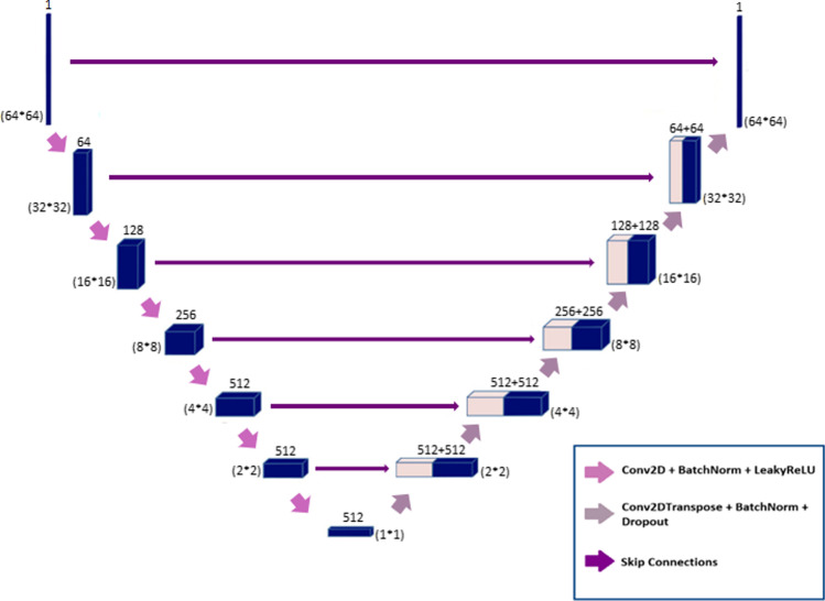
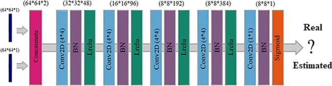
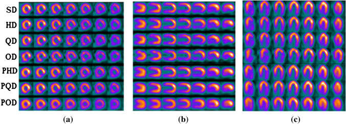
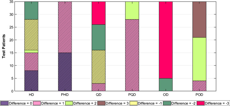
Clinical SPECT-MPI images of 345 patients acquired on a dedicated cardiac SPECT camera in list-mode format were retrospectively employed to predict standard-dose from low-dose images at half-, quarter-, and one-eighth-dose levels. To simulate realistic low-dose projections, 50%, 25%, and 12.5% of the events were randomly selected from the list-mode data through applying binomial subsampling. A generative adversarial network was implemented to predict non-gated standard-dose SPECT images in the projection space at the different dose reduction levels. Well-established metrics, including peak signal-to-noise ratio (PSNR), root mean square error (RMSE), and structural similarity index metrics (SSIM) in addition to Pearson correlation coefficient analysis and clinical parameters derived from Cedars-Sinai software were used to quantitatively assess the predicted standard-dose images. For clinical evaluation, the quality of the predicted standard-dose images was evaluated by a nuclear medicine specialist using a seven-point (- 3 to + 3) grading scheme.
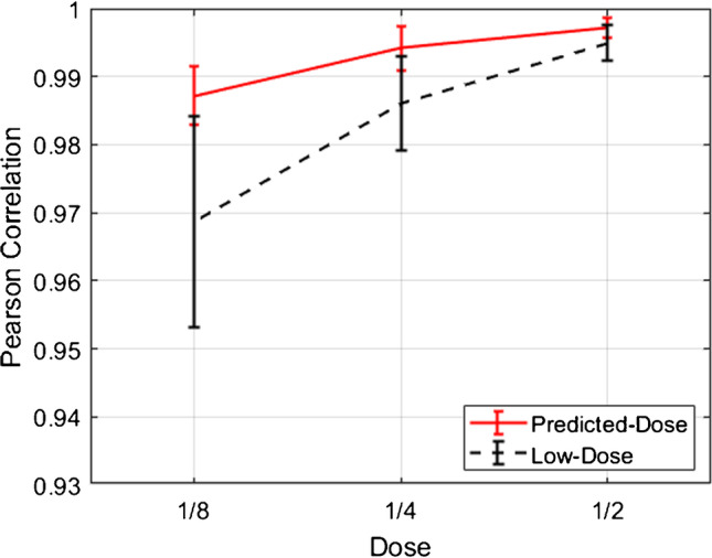
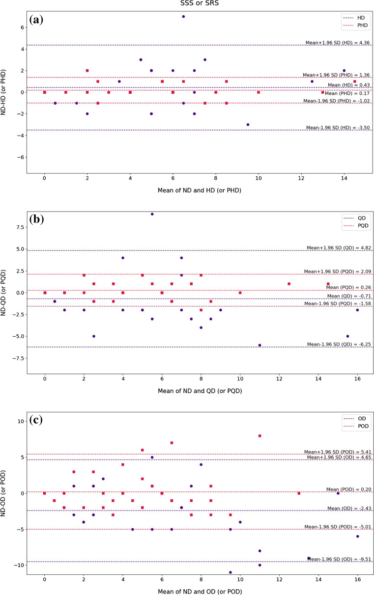
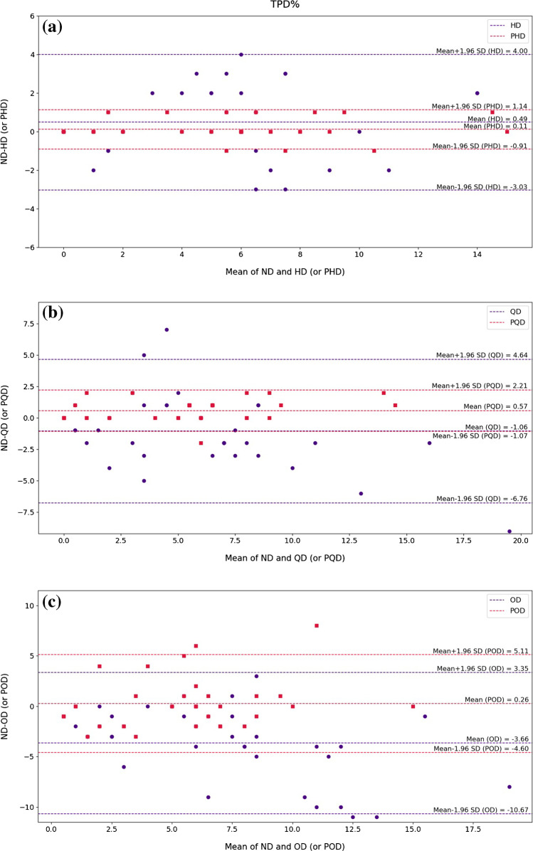
The highest PSNR (42.49 ± 2.37) and SSIM (0.99 ± 0.01) and the lowest RMSE (1.99 ± 0.63) were achieved at a half-dose level. Pearson correlation coefficients were 0.997 ± 0.001, 0.994 ± 0.003, and 0.987 ± 0.004 for the predicted standard-dose images at half-, quarter-, and one-eighth-dose levels, respectively. Using the standard-dose images as reference, the Bland-Altman plots sketched for the Cedars-Sinai selected parameters exhibited remarkably less bias and variance in the predicted standard-dose images compared with the low-dose images at all reduced dose levels. Overall, considering the clinical assessment performed by a nuclear medicine specialist, 100%, 80%, and 11% of the predicted standard-dose images were clinically acceptable at half-, quarter-, and one-eighth-dose levels, respectively.
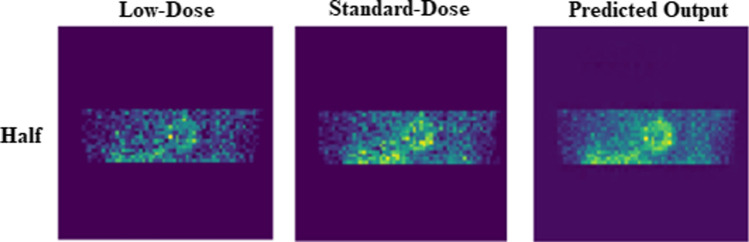
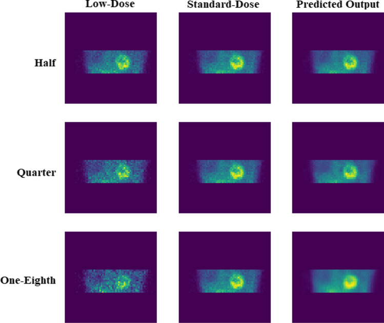
The noise was effectively suppressed by the proposed network, and the predicted standard-dose images were comparable to reference standard-dose images at half- and quarter-dose levels. However, recovery of the underlying signals/information in low-dose images beyond a quarter of the standard dose would not be feasible (due to very poor signal-to-noise ratio) which will adversely affect the clinical interpretation of the resulting images.
本研究旨在探讨在不牺牲诊断准确性的情况下,降低单光子发射型计算机断层心肌灌注成像(SPECT MPI)剂量的可行性。提出了一种深度学习方法,通过在投影空间中不同剂量降低水平上,从相应的低剂量图像中合成全剂量图像。
回顾性地使用专用心脏 SPECT 相机以列表模式格式采集 345 例患者的临床 SPECT-MPI 图像,用于预测半剂量、四分之一剂量和八分之一剂量水平的低剂量图像的标准剂量。为了模拟真实的低剂量投影,通过应用二项式抽样,从列表模式数据中随机选择 50%、25%和 12.5%的事件。实现了生成对抗网络,以预测不同剂量降低水平下投影空间中的非门控标准剂量 SPECT 图像。使用包括峰值信噪比(PSNR)、均方根误差(RMSE)和结构相似性指数度量(SSIM)在内的既定指标,以及来自 Cedars-Sinai 软件的临床参数,对预测的标准剂量图像进行定量评估。为了临床评估,核医学专家使用七点(-3 到+3)评分方案对预测的标准剂量图像的质量进行评估。
在半剂量水平下,获得了最高的 PSNR(42.49±2.37)、SSIM(0.99±0.01)和最低的 RMSE(1.99±0.63)。预测的半剂量、四分之一剂量和八分之一剂量水平的标准剂量图像的皮尔逊相关系数分别为 0.997±0.001、0.994±0.003 和 0.987±0.004。与所有降低剂量水平的低剂量图像相比,使用标准剂量图像作为参考,在预测的标准剂量图像中绘制的 Cedars-Sinai 选择参数的 Bland-Altman 图显示出显著更小的偏差和方差。总体而言,考虑到核医学专家进行的临床评估,在半剂量、四分之一剂量和八分之一剂量水平下,分别有 100%、80%和 11%的预测标准剂量图像在临床上是可以接受的。
所提出的网络有效地抑制了噪声,并且预测的标准剂量图像与半剂量和四分之一剂量水平的参考标准剂量图像相当。然而,在标准剂量的四分之一以下,恢复低剂量图像中的潜在信号/信息将是不可行的(由于信噪比非常差),这将对图像的临床解释产生不利影响。