Neuroscience Axis, Research Center of CHU de Québec - Université Laval, Quebec City, QC, Canada; Department of Psychiatry and Neuroscience, Faculty of Medicine, Université Laval, Quebec City, QC, Canada.
Neuroscience Axis, Research Center of CHU de Québec - Université Laval, Quebec City, QC, Canada; Research Center CERVO, Quebec City, QC, Canada; Department of Psychiatry and Neuroscience, Faculty of Medicine, Université Laval, Quebec City, QC, Canada.
Neurobiol Dis. 2021 Dec;161:105561. doi: 10.1016/j.nbd.2021.105561. Epub 2021 Nov 13.
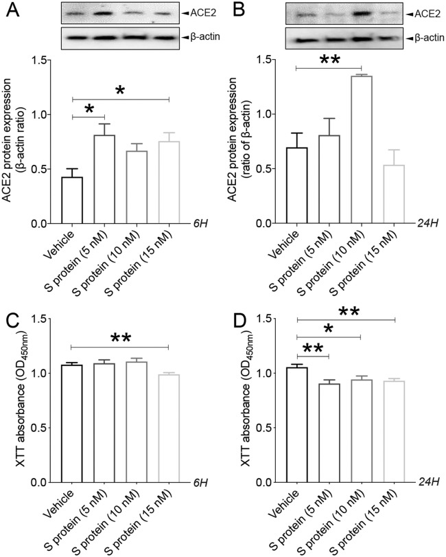
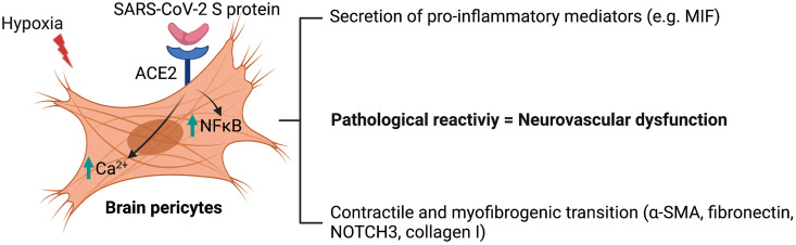
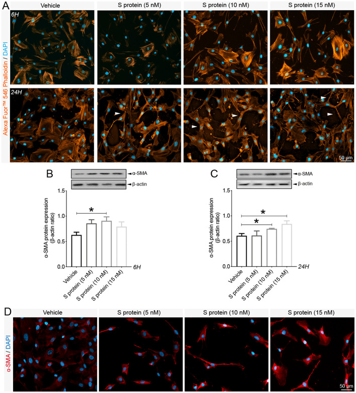
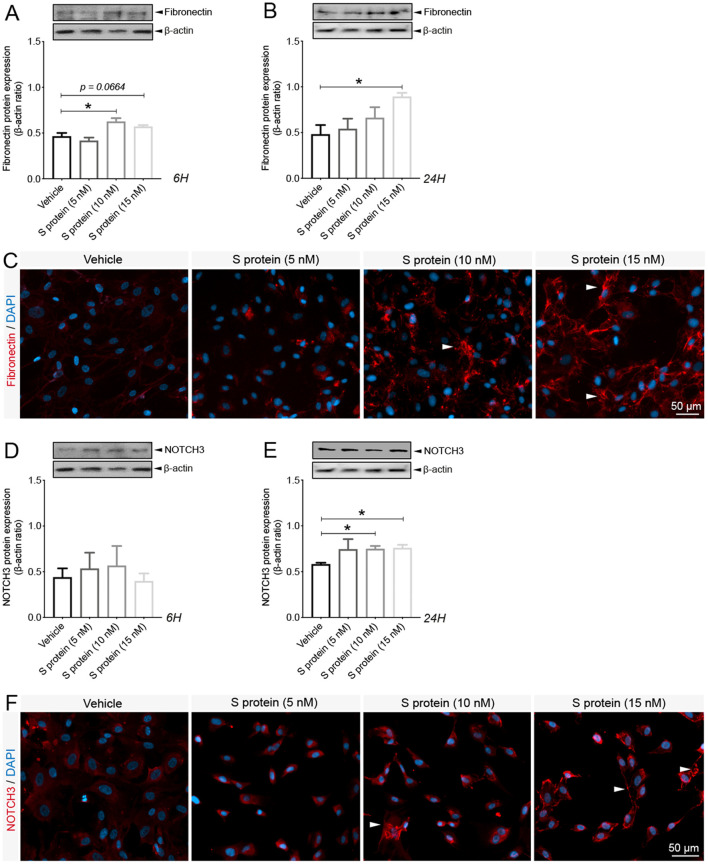
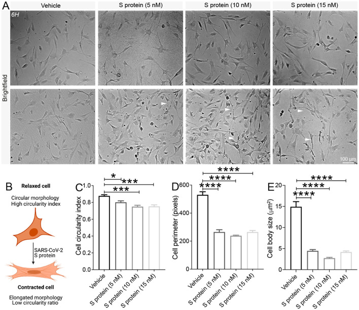
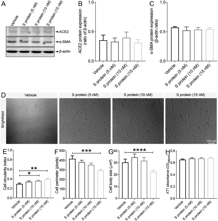
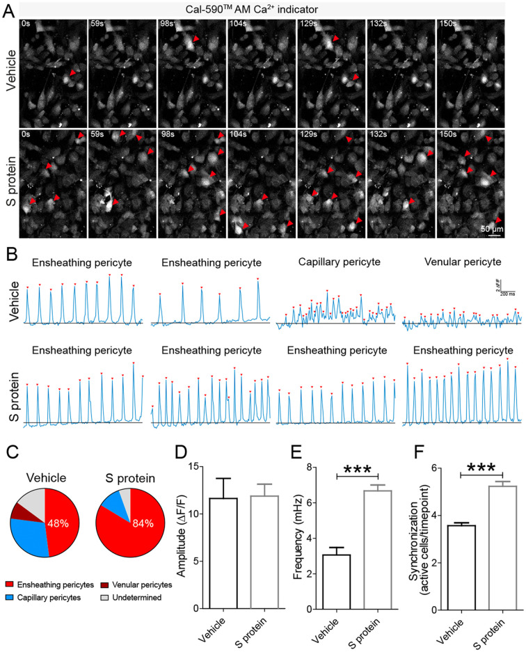
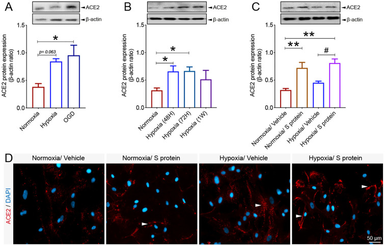
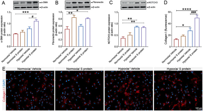
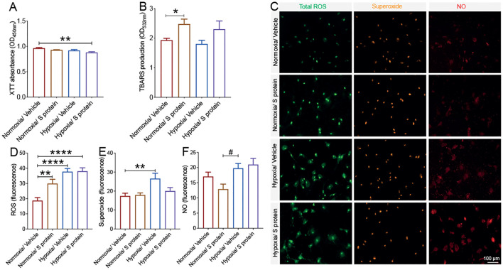
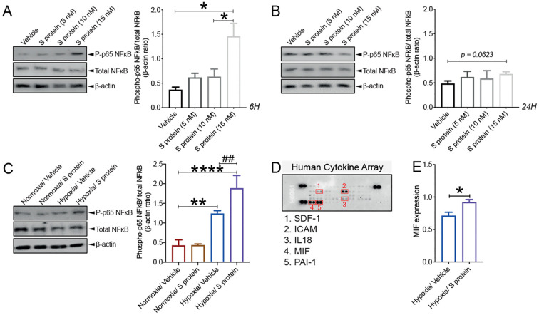
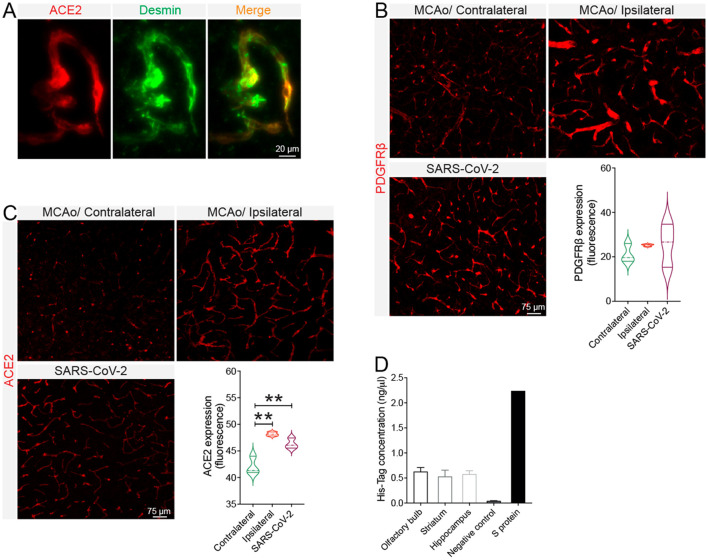
Coronavirus disease 19 (COVID-19) is a respiratory illness caused by severe acute respiratory syndrome coronavirus-2 (SARS-CoV-2). COVID-19 pathogenesis causes vascular-mediated neurological disorders via elusive mechanisms. SARS-CoV-2 infects host cells via the binding of viral Spike (S) protein to transmembrane receptor, angiotensin-converting enzyme 2 (ACE2). Although brain pericytes were recently shown to abundantly express ACE2 at the neurovascular interface, their response to SARS-CoV-2 S protein is still to be elucidated. Using cell-based assays, we found that ACE2 expression in human brain vascular pericytes was increased upon S protein exposure. Pericytes exposed to S protein underwent profound phenotypic changes associated with an elongated and contracted morphology accompanied with an enhanced expression of contractile and myofibrogenic proteins, such as α-smooth muscle actin (α-SMA), fibronectin, collagen I, and neurogenic locus notch homolog protein-3 (NOTCH3). On the functional level, S protein exposure promoted the acquisition of calcium (Ca signature of contractile ensheathing pericytes characterized by highly regular oscillatory Ca fluctuations. Furthermore, S protein induced lipid peroxidation, oxidative and nitrosative stress in pericytes as well as triggered an immune reaction translated by activation of nuclear factor-kappa-B (NF-κB) signaling pathway, which was potentiated by hypoxia, a condition associated with vascular comorbidities that exacerbate COVID-19 pathogenesis. S protein exposure combined to hypoxia enhanced the production of pro-inflammatory cytokines involved in immune cell activation and trafficking, namely macrophage migration inhibitory factor (MIF). Using transgenic mice expressing the human ACE2 that recognizes S protein, we observed that the intranasal infection with SARS-CoV-2 rapidly induced hypoxic/ischemic-like pericyte reactivity in the brain of transgenic mice, accompanied with an increased vascular expression of ACE2. Moreover, we found that SARS-CoV-2 S protein accumulated in the intranasal cavity reached the brain of mice in which the nasal mucosa is deregulated. Collectively, these findings suggest that SARS-CoV-2 S protein impairs the vascular and immune regulatory functions of brain pericytes, which may account for vascular-mediated brain damage. Our study provides a better understanding for the mechanisms underlying cerebrovascular disorders in COVID-19, paving the way to develop new therapeutic interventions.
新型冠状病毒病(COVID-19)是由严重急性呼吸系统综合征冠状病毒 2(SARS-CoV-2)引起的呼吸道疾病。COVID-19 发病机制通过难以捉摸的机制导致血管介导的神经紊乱。SARS-CoV-2 通过病毒刺突(S)蛋白与跨膜受体血管紧张素转换酶 2(ACE2)的结合感染宿主细胞。尽管最近发现脑周细胞在神经血管界面大量表达 ACE2,但它们对 SARS-CoV-2 S 蛋白的反应仍有待阐明。使用基于细胞的测定法,我们发现 S 蛋白暴露会增加人脑血管周细胞中的 ACE2 表达。暴露于 S 蛋白的周细胞经历了与伸长和收缩形态相关的深刻表型变化,同时伴随着收缩和肌成纤维蛋白的表达增强,如α-平滑肌肌动蛋白(α-SMA)、纤维连接蛋白、I 型胶原和神经源性巢蛋白 Notch 同源物 3(NOTCH3)。在功能水平上,S 蛋白暴露促进了钙(Ca)摄取。收缩性鞘周细胞的特征是具有高度规则的振荡性 Ca 波动的钙特征。此外,S 蛋白诱导周细胞脂质过氧化、氧化和硝化应激,以及激活核因子-κB(NF-κB)信号通路引发免疫反应,缺氧会增强 NF-κB 信号通路的激活,缺氧是与血管合并症相关的一种加重 COVID-19 发病机制的情况。S 蛋白暴露与缺氧结合可增强参与免疫细胞激活和迁移的促炎细胞因子的产生,即巨噬细胞移动抑制因子(MIF)。使用表达识别 S 蛋白的人 ACE2 的转基因小鼠,我们观察到 SARS-CoV-2 鼻腔感染迅速诱导转基因小鼠大脑中缺氧/缺血样周细胞反应,同时血管 ACE2 表达增加。此外,我们发现鼻腔腔中积累的 SARS-CoV-2 S 蛋白到达了鼻黏膜失调的小鼠的大脑中。综上所述,这些发现表明 SARS-CoV-2 S 蛋白损害了脑周细胞的血管和免疫调节功能,这可能是血管介导的脑损伤的原因。我们的研究为 COVID-19 中脑血管疾病的发病机制提供了更好的理解,为开发新的治疗干预措施铺平了道路。