• 文献检索
  • 文档翻译
  • 深度研究
  • 学术资讯
  • Suppr Zotero 插件Zotero 插件
  • 邀请有礼
  • 套餐&价格
  • 历史记录
应用&插件
Suppr Zotero 插件Zotero 插件浏览器插件Mac 客户端Windows 客户端微信小程序
定价
高级版会员购买积分包购买API积分包
服务
文献检索文档翻译深度研究API 文档MCP 服务
关于我们
关于 Suppr公司介绍联系我们用户协议隐私条款
关注我们

Suppr 超能文献

核心技术专利:CN118964589B侵权必究
粤ICP备2023148730 号-1Suppr @ 2026

文献检索

告别复杂PubMed语法,用中文像聊天一样搜索,搜遍4000万医学文献。AI智能推荐,让科研检索更轻松。

立即免费搜索

文件翻译

保留排版,准确专业,支持PDF/Word/PPT等文件格式,支持 12+语言互译。

免费翻译文档

深度研究

AI帮你快速写综述,25分钟生成高质量综述,智能提取关键信息,辅助科研写作。

立即免费体验

HA-g-CS 植入物联合中等强度运动刺激软骨下骨重塑,促进小鼠骨软骨缺损修复。

HA-g-CS Implant and Moderate-intensity Exercise Stimulate Subchondral Bone Remodeling and Promote Repair of Osteochondral Defects in Mice.

机构信息

Department of Orthopaedics, Nanfang Hospital, Southern Medical University, Guangzhou, Guangdong 510515, China.

Key Laboratory of Bone and Cartilage Regeneration Medicine, Southern Medical University, Guangzhou, Guangdong 510515, China.

出版信息

Int J Med Sci. 2021 Oct 22;18(16):3808-3820. doi: 10.7150/ijms.63401. eCollection 2021.

DOI:10.7150/ijms.63401
PMID:34790057
原文链接:https://pmc.ncbi.nlm.nih.gov/articles/PMC8579292/
Abstract

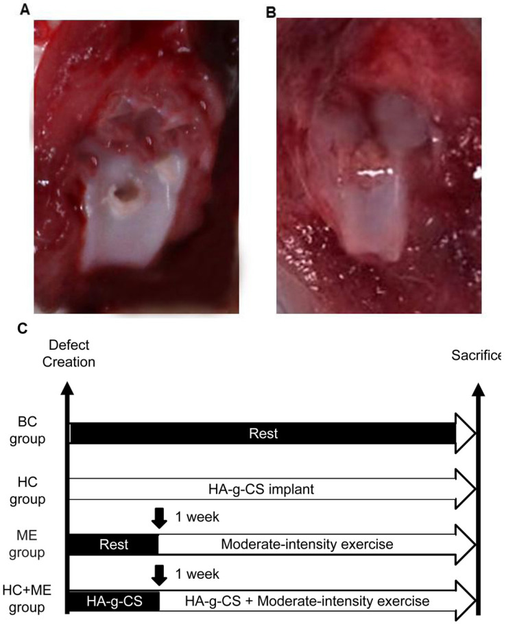
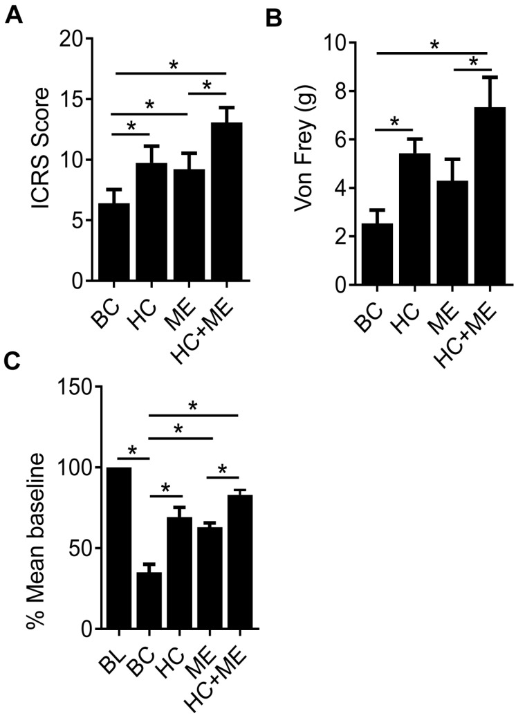
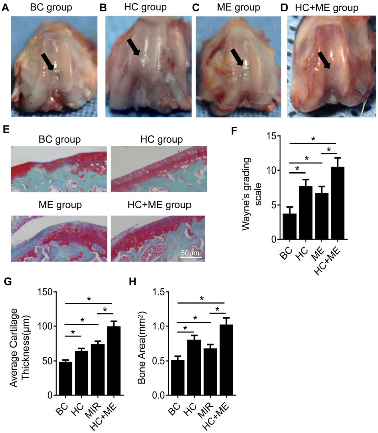
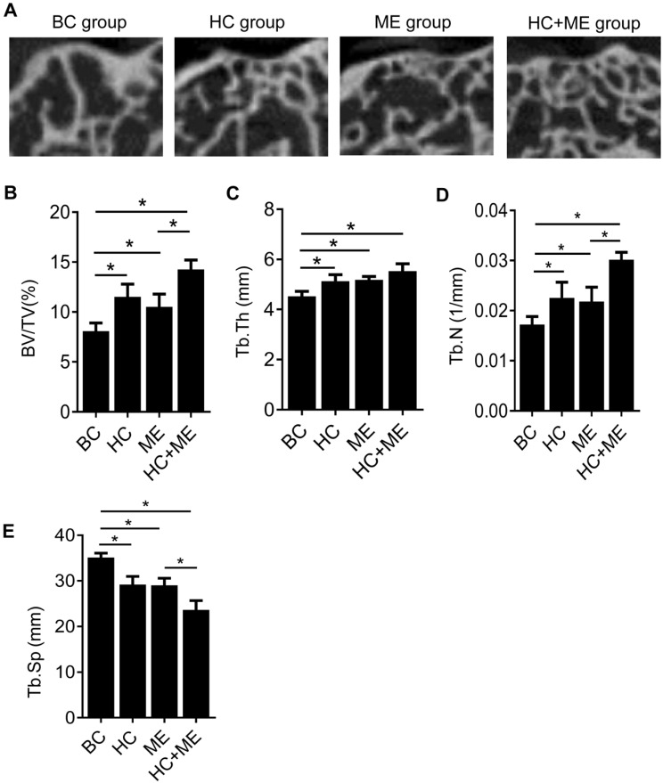
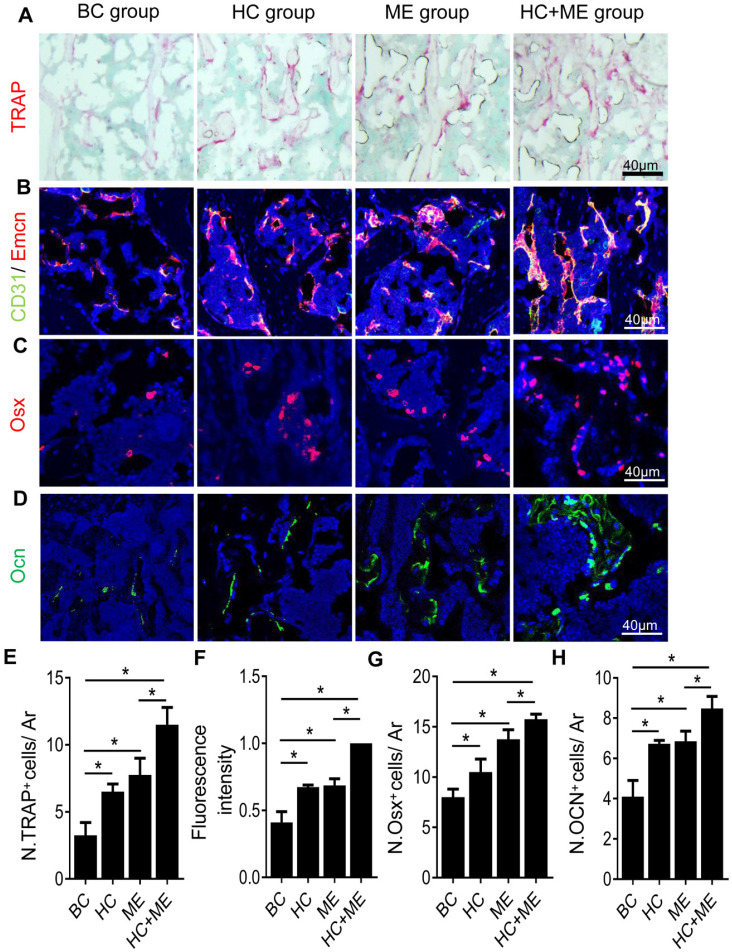
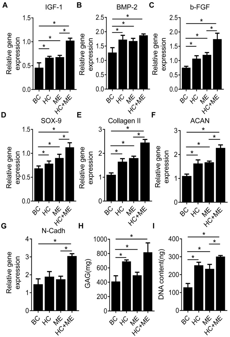
Substantial evidence shows that crosstalk between cartilage and subchondral bone may play an important role in cartilage repair. Animal models have shown that hydroxyapatite-grafted-chitosan implant (HA-g-CS) and moderate-intensity exercise promote regeneration of osteochondral defects. However, no studies have demonstrated that these two factors may have a synergistic activity to facilitate subchondral bone remodeling in mice, thus supporting bone-cartilage repair. This study was to clarify whether HA-g-CS and moderate-intensity exercise might have a synergistic effect on facilitating (1) regeneration of osteochondral defects and (2) subchondral bone remodeling in a mouse model of osteochondral defects. Mouse models of osteochondral defects were created and divided into four groups. BC Group was subjected to no treatment, HC Group to HA-g-CS implantation into osteochondral defects, ME group to moderate-intensity treadmill running exercise, and HC+ME group to both HA-g-CS implantation and moderate-intensity exercise until sacrifice. Extent of subchondral bone remodeling at the injury site and subsequent cartilage repair were assessed at 4 weeks after surgery. Compared with BC group, HC, ME and HC+ME groups showed more cartilage repair and thicker articular cartilage layers and HC+ME group acquired the best results. The extent of cartilage repair was correlated positively to bone formation activity at the injured site as verified by microCT and correlation analysis. Histology and immunofluorescence staining confirmed that bone remodeling activity was increased in HC and ME groups, and especially in HC+ME group. This bone formation process was accompanied by an increase in osteogenesis and chondrogenesis factors at the injury site which promoted cartilage repair. In a mouse model of osteochondral repair, HA-g-CS implant and moderate-intensity exercise may have a synergistic effect on improving osteochondral repair potentially through promotion of subchondral bone remodeling and generation of osteogenesis and chondrogenesis factors. Combination of HA-g-CS implantation and moderate-intensity exercise may be considered potentially in clinic to promote osteochondral defect repair. Also, cartilage and subchondral bone forms a functional unit in an articular joint and subchondral bone may regulate cartilage repair by secreting growth factors in its remodeling process. However, a deeper insight into the exact role of HA-g-CS implantation and moderate-intensity exercise in promoting osteochondral repair in other animal models should be explored before they can be applied in clinic in the future.

摘要

大量证据表明,软骨和软骨下骨之间的串扰可能在软骨修复中发挥重要作用。动物模型表明,羟基磷灰石接枝壳聚糖植入物(HA-g-CS)和中等强度运动促进了骨软骨缺损的再生。然而,尚无研究表明这两个因素可能具有协同作用,从而促进小鼠软骨下骨重塑,从而支持骨-软骨修复。本研究旨在阐明 HA-g-CS 和中等强度运动是否可能对促进(1)骨软骨缺损的再生和(2)骨软骨缺损小鼠模型中的软骨下骨重塑具有协同作用。创建了骨软骨缺损的小鼠模型,并将其分为四组。BC 组未进行任何治疗,HC 组将 HA-g-CS 植入骨软骨缺损,ME 组进行中等强度跑步机运动,HC+ME 组进行 HA-g-CS 植入和中等强度运动,直至处死。手术后 4 周评估损伤部位软骨下骨重塑的程度和随后的软骨修复情况。与 BC 组相比,HC、ME 和 HC+ME 组显示出更多的软骨修复和更厚的关节软骨层,而 HC+ME 组的效果最佳。软骨修复的程度与受伤部位的骨形成活性呈正相关,这一点通过 microCT 和相关分析得到了验证。组织学和免疫荧光染色证实,HC 和 ME 组骨重塑活性增加,尤其是在 HC+ME 组。这种骨形成过程伴随着损伤部位成骨和成软骨因子的增加,从而促进了软骨修复。在骨软骨修复的小鼠模型中,HA-g-CS 植入物和中等强度运动可能通过促进软骨下骨重塑和产生成骨和成软骨因子,对改善骨软骨修复具有协同作用。HA-g-CS 植入物和中等强度运动的联合应用可能在临床上被考虑用于促进骨软骨缺损修复。此外,软骨和软骨下骨在关节中形成一个功能单元,软骨下骨在其重塑过程中通过分泌生长因子来调节软骨修复。然而,在将来将其应用于临床之前,应该在其他动物模型中更深入地探讨 HA-g-CS 植入物和中等强度运动在促进骨软骨修复中的确切作用。

https://cdn.ncbi.nlm.nih.gov/pmc/blobs/f47d/8579292/32578cd9b6ea/ijmsv18p3808g006.jpg
https://cdn.ncbi.nlm.nih.gov/pmc/blobs/f47d/8579292/1e51ce93a1e1/ijmsv18p3808g001.jpg
https://cdn.ncbi.nlm.nih.gov/pmc/blobs/f47d/8579292/21c74e02cc92/ijmsv18p3808g002.jpg
https://cdn.ncbi.nlm.nih.gov/pmc/blobs/f47d/8579292/43985130754d/ijmsv18p3808g003.jpg
https://cdn.ncbi.nlm.nih.gov/pmc/blobs/f47d/8579292/19990ba9e0be/ijmsv18p3808g004.jpg
https://cdn.ncbi.nlm.nih.gov/pmc/blobs/f47d/8579292/a4dad562939c/ijmsv18p3808g005.jpg
https://cdn.ncbi.nlm.nih.gov/pmc/blobs/f47d/8579292/32578cd9b6ea/ijmsv18p3808g006.jpg

相似文献

1
HA-g-CS Implant and Moderate-intensity Exercise Stimulate Subchondral Bone Remodeling and Promote Repair of Osteochondral Defects in Mice.HA-g-CS 植入物联合中等强度运动刺激软骨下骨重塑,促进小鼠骨软骨缺损修复。
Int J Med Sci. 2021 Oct 22;18(16):3808-3820. doi: 10.7150/ijms.63401. eCollection 2021.
2
Preparation of a biphase composite scaffold and its application in tissue engineering for femoral osteochondral defects in rabbits.双相复合支架的制备及其在兔股骨骨软骨缺损组织工程中的应用。
Int Orthop. 2017 Sep;41(9):1899-1908. doi: 10.1007/s00264-017-3522-2. Epub 2017 Jun 14.
3
Osteochondral repair using a scaffold-free tissue-engineered construct derived from synovial mesenchymal stem cells and a hydroxyapatite-based artificial bone.使用源自滑膜间充质干细胞和羟基磷灰石基人工骨的无支架组织工程构建体进行骨软骨修复。
Tissue Eng Part A. 2014 Sep;20(17-18):2291-304. doi: 10.1089/ten.tea.2013.0414. Epub 2014 Mar 21.
4
[Cartilage repair and subchondral bone reconstruction based on three-dimensional printing technique].基于三维打印技术的软骨修复与软骨下骨重建
Zhongguo Xiu Fu Chong Jian Wai Ke Za Zhi. 2014 Mar;28(3):318-24.
5
A novel, self-assembled artificial cartilage-hydroxyapatite conjugate for combined articular cartilage and subchondral bone repair: histopathological analysis of cartilage tissue engineering in rat knee joints.一种新型自组装人工软骨-羟基磷灰石缀合物用于关节软骨和软骨下骨联合修复:大鼠膝关节软骨组织工程的组织病理学分析。
Int J Nanomedicine. 2019 Feb 19;14:1283-1298. doi: 10.2147/IJN.S193963. eCollection 2019.
6
Cell-free multi-layered collagen-based scaffolds demonstrate layer specific regeneration of functional osteochondral tissue in caprine joints.无细胞多层胶原基支架在山羊关节中显示出功能性骨软骨组织的层特异性再生。
Biomaterials. 2016 May;87:69-81. doi: 10.1016/j.biomaterials.2016.02.006. Epub 2016 Feb 9.
7
A single integrated osteochondral in situ composite scaffold with a multi-layered functional structure.具有多层次功能结构的一体化原位复合骨软骨支架
Colloids Surf B Biointerfaces. 2018 Jul 1;167:354-363. doi: 10.1016/j.colsurfb.2018.04.029. Epub 2018 Apr 16.
8
3D printing of fibre-reinforced cartilaginous templates for the regeneration of osteochondral defects.用于骨软骨缺损再生的纤维增强软骨模板的3D打印
Acta Biomater. 2020 Sep 1;113:130-143. doi: 10.1016/j.actbio.2020.05.040. Epub 2020 Jun 4.
9
[RELATIONSHIP BETWEEN SUBCHONDRAL BONE RECONSTRUCTION AND ARTICULAR CARTILAGE REGENERATION IN A RABBIT MODEL OF SPONTANEOUS OSTEOCHONDRAL REPAIR].[兔自发性骨软骨修复模型中软骨下骨重建与关节软骨再生的关系]
Zhongguo Xiu Fu Chong Jian Wai Ke Za Zhi. 2014 Jun;28(6):681-6.
10
Novel hydroxyapatite/chitosan bilayered scaffold for osteochondral tissue-engineering applications: Scaffold design and its performance when seeded with goat bone marrow stromal cells.用于骨软骨组织工程应用的新型羟基磷灰石/壳聚糖双层支架:支架设计及其接种山羊骨髓基质细胞后的性能。
Biomaterials. 2006 Dec;27(36):6123-37. doi: 10.1016/j.biomaterials.2006.07.034. Epub 2006 Aug 30.

引用本文的文献

1
dynamic visualization and evaluation of collagen degradation utilizing NIR-II fluorescence imaging in mice models.利用近红外二区荧光成像在小鼠模型中对胶原蛋白降解进行动态可视化和评估。
Regen Biomater. 2025 Apr 11;12:rbaf025. doi: 10.1093/rb/rbaf025. eCollection 2025.
2
Hybrid Nanoparticle Engineered with Transforming Growth Factor -β1-Overexpressed Extracellular Vesicle and Cartilage-Targeted Anti-Inflammatory Liposome for Osteoarthritis.用转化生长因子-β1过表达的细胞外囊泡和软骨靶向抗炎脂质体工程化的杂合纳米颗粒用于骨关节炎
ACS Nano. 2024 Dec 17;18(50):33937-33952. doi: 10.1021/acsnano.4c07992. Epub 2024 Dec 8.
3

本文引用的文献

1
Evaluation of an Autologous Bone Mesenchymal Stem Cell-Derived Extracellular Matrix Scaffold in a Rabbit and Minipig Model of Cartilage Repair.评价一种自体骨髓间充质干细胞衍生细胞外基质支架在兔和小型猪关节软骨修复模型中的应用。
Med Sci Monit. 2019 Sep 30;25:7342-7350. doi: 10.12659/MSM.916481.
2
Intra-articularly injected mesenchymal stem cells promote cartilage regeneration, but do not permanently engraft in distant organs.关节内注射间充质干细胞可促进软骨再生,但不会在远处器官内永久定植。
Sci Rep. 2019 Jul 12;9(1):10153. doi: 10.1038/s41598-019-46554-5.
3
Treatment of Articular Cartilage Defects: Focus on Tissue Engineering.
Moderate mechanical stress suppresses chondrocyte ferroptosis in osteoarthritis by regulating NF-κB p65/GPX4 signaling pathway.
适度机械应激通过调控 NF-κB p65/GPX4 信号通路抑制骨关节炎软骨细胞铁死亡。
Sci Rep. 2024 Mar 1;14(1):5078. doi: 10.1038/s41598-024-55629-x.
4
A Review of Cartilage Defect Treatments Using Chitosan Hydrogels in Experimental Animal Models.壳聚糖水凝胶在实验动物模型中治疗软骨缺损的研究进展。
Curr Pharm Biotechnol. 2024;25(8):1058-1072. doi: 10.2174/0113892010245946230919062908.
5
Costal Cartilage Graft Repair Osteochondral Defect in a Mouse Model.肋软骨移植修复小鼠模型中的骨软骨缺损
Cartilage. 2025 Jun;16(2):212-223. doi: 10.1177/19476035231209404. Epub 2023 Oct 26.
6
Crosstalk between Bone and Muscles during Physical Activity.体力活动中骨骼与肌肉的相互作用。
Cells. 2023 Aug 18;12(16):2088. doi: 10.3390/cells12162088.
7
Osteoarthritis animal models for biomaterial-assisted osteochondral regeneration.用于生物材料辅助骨软骨再生的骨关节炎动物模型
Biomater Transl. 2022 Dec 28;3(4):264-279. doi: 10.12336/biomatertransl.2022.04.006. eCollection 2022.
8
Icariin-conditioned serum combined with chitosan attenuates cartilage injury in rabbit knees with osteochondral defect.淫羊藿苷条件培养液联合壳聚糖减轻兔膝关节骨软骨缺损模型的软骨损伤
J Orthop Surg Res. 2023 Feb 21;18(1):125. doi: 10.1186/s13018-023-03607-w.
关节软骨缺损的治疗:聚焦于组织工程
In Vivo. 2018 Nov-Dec;32(6):1289-1300. doi: 10.21873/invivo.11379.
4
Molecular dynamics simulations of adsorption and desorption of bone morphogenetic protein-2 on textured hydroxyapatite surfaces.骨形态发生蛋白-2 在纹理化羟基磷灰石表面吸附和解吸的分子动力学模拟。
Acta Biomater. 2018 Oct 15;80:121-130. doi: 10.1016/j.actbio.2018.09.019. Epub 2018 Sep 15.
5
The Importance of Subchondral Bone in the Pathophysiology of Osteoarthritis.软骨下骨在骨关节炎病理生理学中的重要性。
Front Vet Sci. 2018 Aug 28;5:178. doi: 10.3389/fvets.2018.00178. eCollection 2018.
6
Upregulation of SIRT1 by Kartogenin Enhances Antioxidant Functions and Promotes Osteogenesis in Human Mesenchymal Stem Cells.软骨素糖胺通过上调 SIRT1 增强人骨髓间充质干细胞的抗氧化功能并促进成骨分化。
Oxid Med Cell Longev. 2018 Jul 15;2018:1368142. doi: 10.1155/2018/1368142. eCollection 2018.
7
Return to Play Among Elite Basketball Players After Osteochondral Allograft Transplantation of Full-Thickness Cartilage Lesions.全层软骨损伤异体骨软骨移植后精英篮球运动员的重返赛场情况
Orthop J Sports Med. 2018 Jul 25;6(7):2325967118786941. doi: 10.1177/2325967118786941. eCollection 2018 Jul.
8
Tailoring the subchondral bone phase of a multi-layered osteochondral construct to support bone healing and a cartilage analog.定制多层骨软骨构建体的软骨下骨相以支持骨愈合和软骨类似物。
Acta Biomater. 2018 Sep 15;78:351-364. doi: 10.1016/j.actbio.2018.08.009. Epub 2018 Aug 10.
9
Early Changes of Articular Cartilage and Subchondral Bone in The DMM Mouse Model of Osteoarthritis.骨关节炎 DMM 小鼠模型中软骨和软骨下骨的早期变化。
Sci Rep. 2018 Feb 12;8(1):2855. doi: 10.1038/s41598-018-21184-5.
10
Positive-Feedback Regulation of Subchondral H-Type Vessel Formation by Chondrocyte Promotes Osteoarthritis Development in Mice.软骨细胞正反馈调控软骨下 H 型血管形成促进小鼠骨关节炎的发生。
J Bone Miner Res. 2018 May;33(5):909-920. doi: 10.1002/jbmr.3388. Epub 2018 Mar 24.