Kramer Ashley C, Gurdziel Katherine, Thummel Ryan
Department of Ophthalmology, Visual and Anatomical Sciences, Wayne State University School of Medicine, Detroit, MI, United States.
Genome Sciences Core, Wayne State University, Detroit, MI, United States.
Front Cell Dev Biol. 2021 Nov 1;9:741514. doi: 10.3389/fcell.2021.741514. eCollection 2021.
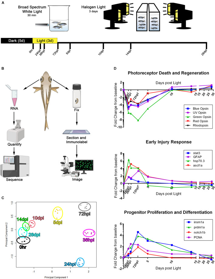
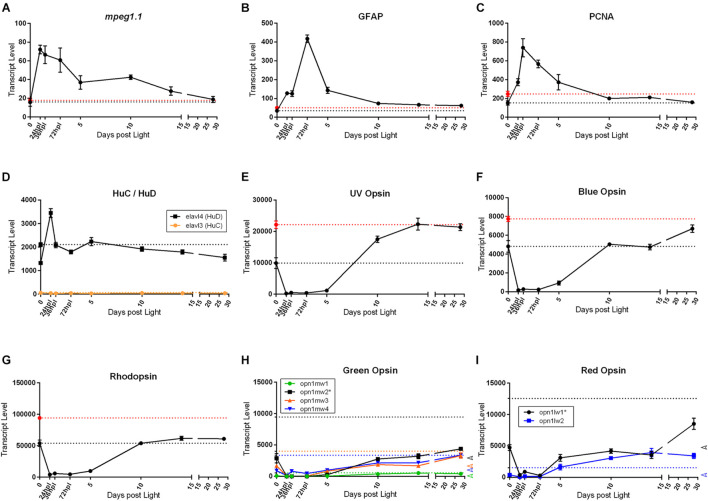
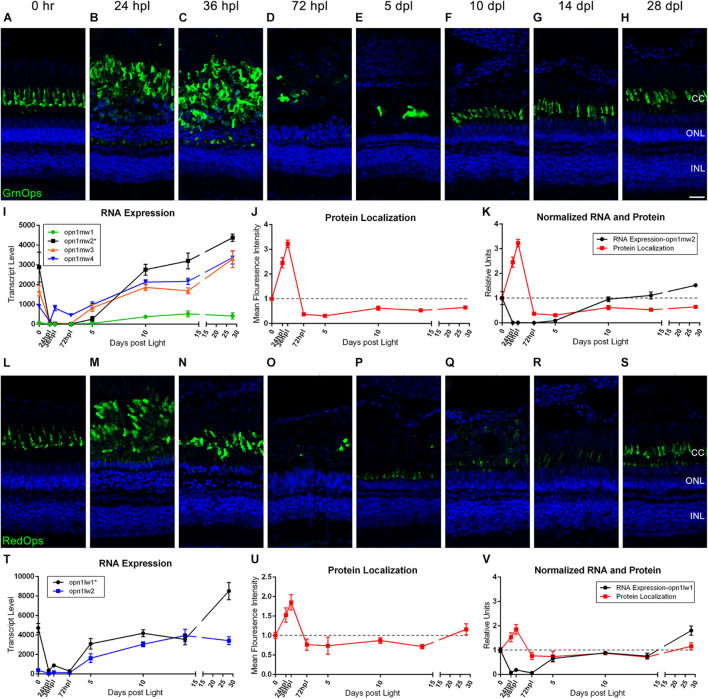
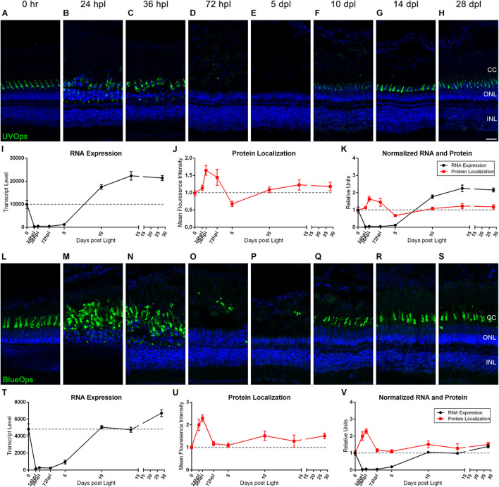
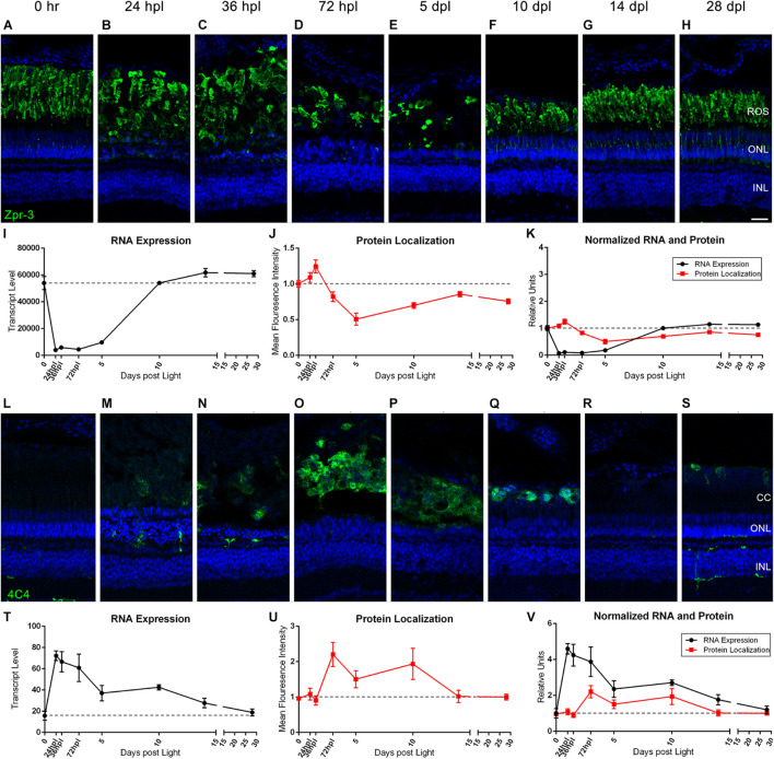
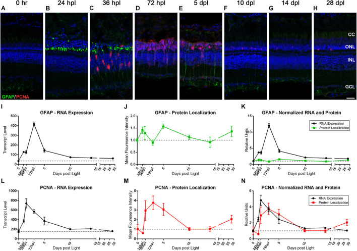
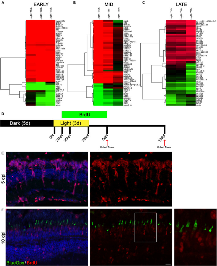
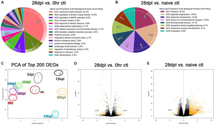
Following photoreceptors ablation by intense light exposure, adult zebrafish are capable of complete regeneration due to the ability of their Müller glia (MG) to re-enter the cell cycle, creating progenitors that differentiate into new photoreceptors. The majority of previous reports on retinal regeneration focused on the first few days of the regenerative response, which include MG cell-cycle re-entry and progenitor cell proliferation. With this study, we analyzed the full 28-day time-course of regeneration by pairing a detailed morphological/immunological analysis with RNA-seq transcriptional profiling at 8 key time points during retinal regeneration. We observed several novel findings. First, we provide evidence for two separate peaks of MG gliosis, with the secondary gliotic peak occurring after MG cell-cycle re-entry. Second, we highlight a distinct transcriptional shift between 5- and 10-days post lesion that highlights the transition from progenitor proliferation to differentiation into new photoreceptors. Third, we show distinctly different patterns of transcriptional recovery of the photoreceptor opsins at 28 days post lesion. Finally, using differential gene expression analysis, we revealed that the established functional recovery of the retina at 28 days post lesion does not, in fact, return to an undamaged transcriptional state, potentially redefining what the field considers complete regeneration. Together, to our knowledge, this work represents the first histological and transcriptomic map of a 28-day time-course of retinal regeneration in adult zebrafish.
在通过强光照射使光感受器消融后,成年斑马鱼能够完全再生,这是因为它们的米勒胶质细胞(MG)具有重新进入细胞周期的能力,从而产生分化为新光感受器的祖细胞。此前关于视网膜再生的大多数报告都集中在再生反应的最初几天,包括MG细胞周期重新进入和祖细胞增殖。在这项研究中,我们通过在视网膜再生的8个关键时间点将详细的形态学/免疫学分析与RNA测序转录谱分析相结合,分析了整个28天的再生时间进程。我们观察到了几个新发现。首先,我们为MG胶质增生的两个独立峰值提供了证据,继发性胶质增生峰值发生在MG细胞周期重新进入之后。其次,我们强调了损伤后5天至10天之间明显的转录转变,这突出了从祖细胞增殖到分化为新光感受器的转变。第三,我们展示了损伤后28天时光感受器视蛋白转录恢复的明显不同模式。最后,通过差异基因表达分析,我们发现损伤后28天视网膜已确立的功能恢复实际上并未恢复到未受损的转录状态,这可能会重新定义该领域所认为的完全再生。据我们所知,这项工作共同代表了成年斑马鱼视网膜再生28天时间进程的首张组织学和转录组图谱。