Department of General, Visceral and Transplantation Surgery, University Hospital, LMU Munich, Munich, Germany.
Department of Medicine III, University Hospital, LMU Munich, Munich, Germany.
BMC Cancer. 2021 Nov 18;21(1):1243. doi: 10.1186/s12885-021-08927-w.
Molecular differences in colorectal cancer (CRC) are associated with the metastatic route. Patient survival is mainly driven by metastatic spread thus it is imperative to understand its key drivers to develop biomarkers for risk stratification, follow-up protocols and personalized therapy. Thus, this study aimed to identify genes associated with the metastatic route in CRC.
CRC patients resected at our clinic from 2005 to 2014 and with a minimum 5-year follow-up were included in this analysis and grouped into CRC with hepatic (HEP), peritoneal (PER) or without distant metastases (M0), and HEP/PER. Firstly, tumor RNA of 6 patients each was isolated by microdissection from formalin-fixed paraffin-embedded specimens and analyzed by a NanoString analysis. Subsequently, these results were validated with immunohistochemistry and correlated to clinicopathological parameters in a larger collective of CRC patients (HEP n = 51, PER n = 44, M0 n = 47, HEP/PER n = 28).
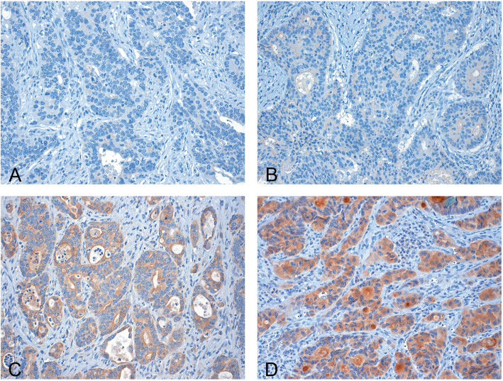
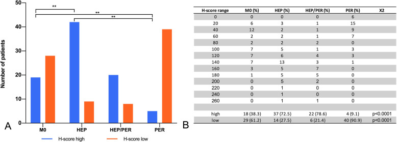
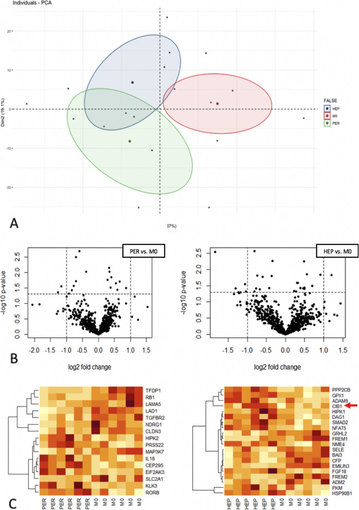
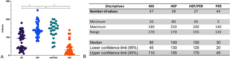
Compared to M0, HEP tumors showed 20 differentially expressed genes associated with epithelial-mesenchymal transition (EMT) and angiogenesis. Compared to M0, PER tumors had 18 differentially expressed genes. The finding of different gene signatures was supported by the multidimensional principal component clustering analysis. Tumor perforation did not influence the metastatic route. CIB1 was homogenously and significantly overexpressed in HEP compared to M0 (p < 0.001), but not in PER. Furthermore, immunohistochemical validation demonstrated that the mean CIB1 expression in HEP was 80% higher than in M0 (p < 0.001).
Gene expression analysis revealed that CIB1 is significantly overexpressed in CRC leading to liver metastases compared to M0 and PER. Thus, the present results suggest that CIB1 may play a crucial role for hematogenous spread to the liver but not for peritoneal carcinomatosis. Consequently, CIB1 seems to be a promising prognostic marker and a potential tool for future targeted therapies as well as early diagnostics and follow-up.
结直肠癌(CRC)的分子差异与转移途径有关。患者的生存主要受转移扩散的影响,因此,了解其关键驱动因素对于开发风险分层、随访方案和个体化治疗的生物标志物至关重要。因此,本研究旨在鉴定与 CRC 转移途径相关的基因。
本研究纳入了 2005 年至 2014 年在我们诊所接受手术切除且随访时间至少 5 年的 CRC 患者,并将其分为肝转移(HEP)、腹膜转移(PER)或无远处转移(M0)和 HEP/PER 组。首先,通过微切割从福尔马林固定石蜡包埋标本中分离出 6 例患者的肿瘤 RNA,并通过 NanoString 分析进行分析。随后,在更大的 CRC 患者群体(HEP n=51、PER n=44、M0 n=47、HEP/PER n=28)中,通过免疫组织化学验证这些结果,并与临床病理参数相关联。
与 M0 相比,HEP 肿瘤显示出 20 个与上皮-间质转化(EMT)和血管生成相关的差异表达基因。与 M0 相比,PER 肿瘤有 18 个差异表达基因。多维主成分聚类分析支持了不同基因特征的发现。肿瘤穿孔并不影响转移途径。与 M0 相比,HEP 中 CIB1 均匀且显著过表达(p<0.001),但在 PER 中没有。此外,免疫组织化学验证表明,HEP 中 CIB1 的平均表达水平比 M0 高 80%(p<0.001)。
基因表达分析显示,与 M0 和 PER 相比,CIB1 在导致肝转移的 CRC 中显著过表达。因此,本研究结果表明,CIB1 可能在血液播散到肝脏中发挥关键作用,但在腹膜转移中不起作用。因此,CIB1 似乎是一种很有前途的预后标志物,也是未来靶向治疗、早期诊断和随访的潜在工具。