Ni Tao, Zhu Yanan, Yang Zhengyi, Xu Chaoyi, Chaban Yuriy, Nesterova Tanya, Ning Jiying, Böcking Till, Parker Michael W, Monnie Christina, Ahn Jinwoo, Perilla Juan R, Zhang Peijun
Division of Structural Biology, Wellcome Trust Centre for Human Genetics, University of Oxford, Oxford OX3 7BN, UK.
Electron Bio-Imaging Centre, Diamond Light Source, Harwell Science and Innovation Campus, Didcot OX11 0DE, UK.
Sci Adv. 2021 Nov 19;7(47):eabj5715. doi: 10.1126/sciadv.abj5715.
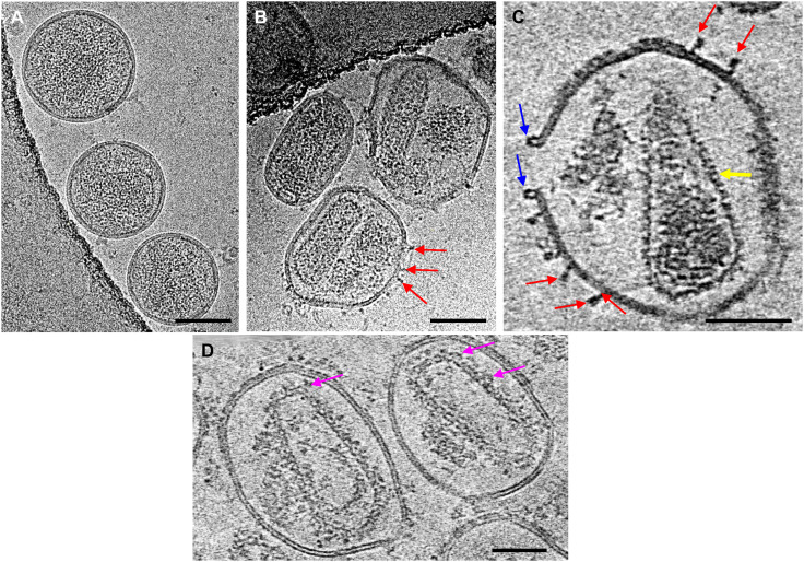
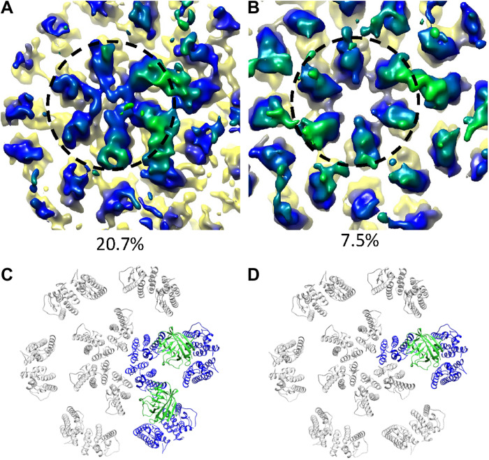
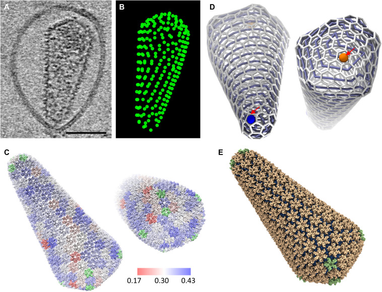
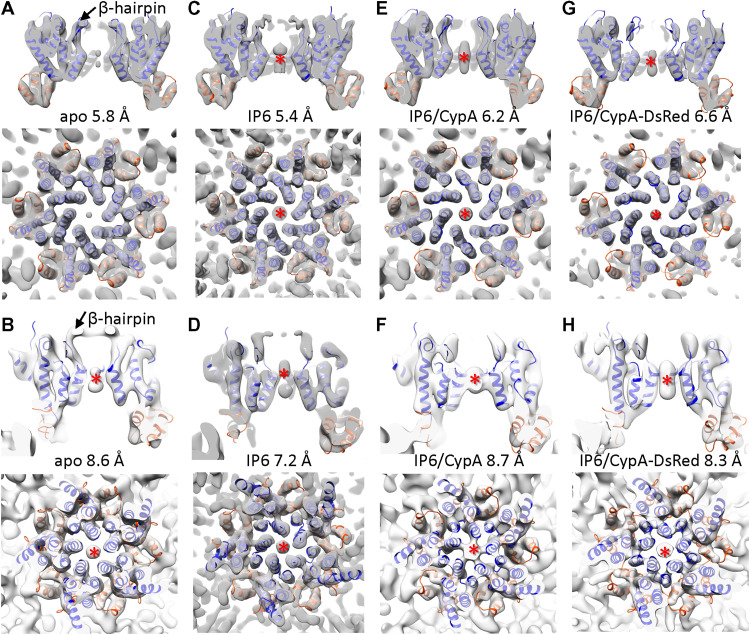
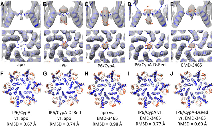
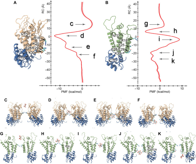
The viral capsid plays essential roles in HIV replication and is a major platform engaging host factors. To overcome challenges in study native capsid structure, we used the perfringolysin O to perforate the membrane of HIV-1 particles, thus allowing host proteins and small molecules to access the native capsid while improving cryo–electron microscopy image quality. Using cryo–electron tomography and subtomogram averaging, we determined the structures of native capsomers in the presence and absence of inositol hexakisphosphate (IP6) and cyclophilin A and constructed an all-atom model of a complete HIV-1 capsid. Our structures reveal two IP6 binding sites and modes of cyclophilin A interactions. Free energy calculations substantiate the two binding sites at R18 and K25 and further show a prohibitive energy barrier for IP6 to pass through the pentamer. Our results demonstrate that perfringolysin O perforation is a valuable tool for structural analyses of enveloped virus capsids and interactions with host cell factors.
病毒衣壳在HIV复制中起着至关重要的作用,是一个与宿主因子相互作用的主要平台。为了克服研究天然衣壳结构时遇到的挑战,我们使用产气荚膜梭菌溶血素O在HIV-1颗粒膜上穿孔,从而使宿主蛋白和小分子能够接触到天然衣壳,同时提高冷冻电子显微镜图像质量。利用冷冻电子断层扫描和亚断层图平均技术,我们确定了在存在和不存在肌醇六磷酸(IP6)及亲环素A的情况下天然衣壳粒的结构,并构建了完整HIV-1衣壳的全原子模型。我们的结构揭示了两个IP6结合位点和亲环素A的相互作用模式。自由能计算证实了R18和K25处的两个结合位点,并进一步表明IP6穿过五聚体存在过高的能量屏障。我们的结果表明,产气荚膜梭菌溶血素O穿孔是用于分析包膜病毒衣壳结构及其与宿主细胞因子相互作用的一种有价值的工具。