PCD R&D Group, Health & Medical Equipment Business Unit, Samsung Electronics, Suwon-si, Gyeonggido, Korea.
Laboratory Animal Center, Daegu Gyeongbuk Medical Innovation Foundation, Daegu, Korea.
Sci Rep. 2021 Nov 23;11(1):22731. doi: 10.1038/s41598-021-02210-5.
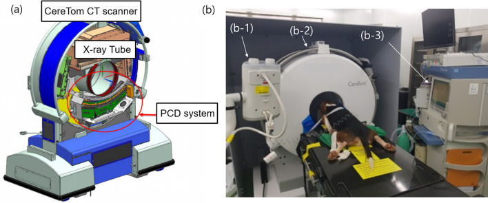
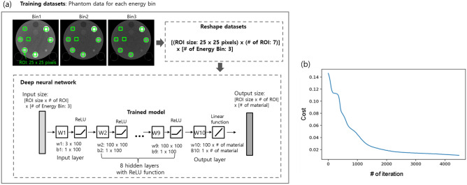
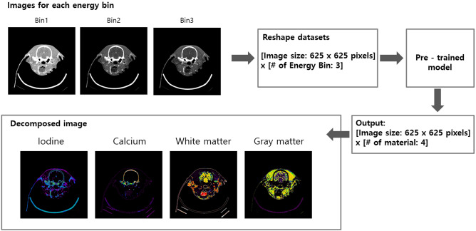
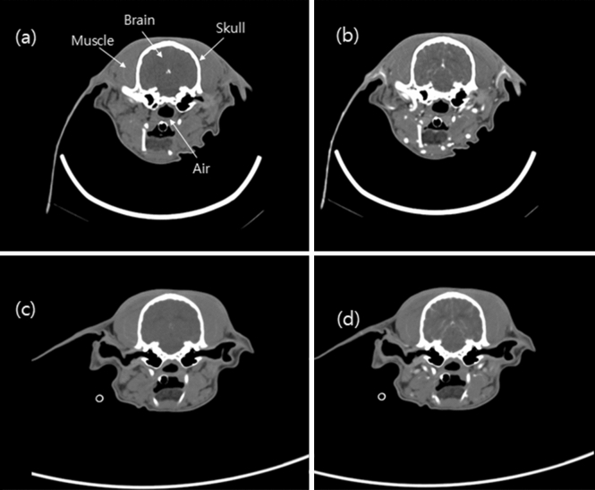
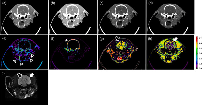
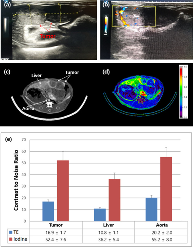
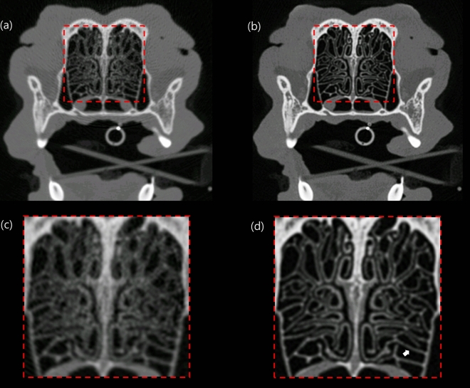
In this study, preclinical experiments were performed with an in-house developed prototypal photon-counting detector computed tomography (PCD CT) system. The performance of the system was compared with the conventional energy-integrating detector (EID)-based CT, concerning the basic image quality biomarkers and the respective capacities for material separation. The pre- and the post-contrast axial images of a canine brain captured by the PCD CT and EID CT systems were found to be visually similar. Multi-energy images were acquired using the PCD CT system, and machine learning-based material decomposition was performed to segment the white and gray matters for the first time in soft tissue segmentation. Furthermore, to accommodate clinical applications that require high resolution acquisitions, a small, native, high-resolution (HR) detector was implemented on the PCD CT system, and its performance was evaluated based on animal experiments. The HR acquisition mode improved the spatial resolution and delineation of the fine structures in the canine's nasal turbinates compared to the standard mode. Clinical applications that rely on high-spatial resolution expectedly will also benefit from this resolution-enhancing function. The results demonstrate the potential impact on the brain tissue segmentation, improved detection of the liver tumors, and capacity to reconstruct high-resolution images both preclinically and clinically.
在这项研究中,使用内部开发的原型光子计数探测器 CT(PCD CT)系统进行了临床前实验。该系统的性能与传统的能量积分探测器(EID)CT 进行了比较,涉及基本的图像质量生物标志物以及各自的材料分离能力。PCD CT 和 EID CT 系统捕获的犬脑的轴向对比前后图像在视觉上相似。使用 PCD CT 系统获取多能量图像,并首次使用基于机器学习的材料分解技术对软组织进行分割,以分割白质和灰质。此外,为了适应需要高分辨率采集的临床应用,在 PCD CT 系统上实现了小型、原生、高分辨率(HR)探测器,并基于动物实验对其性能进行了评估。与标准模式相比,HR 采集模式提高了犬鼻鼻甲的空间分辨率和精细结构的描绘。预计依赖高空间分辨率的临床应用也将受益于这种分辨率增强功能。研究结果表明,该技术在脑组织分割、提高肝脏肿瘤检测以及在临床前和临床中重建高分辨率图像的能力方面具有潜在影响。