• 文献检索
  • 文档翻译
  • 深度研究
  • 学术资讯
  • Suppr Zotero 插件Zotero 插件
  • 邀请有礼
  • 套餐&价格
  • 历史记录
应用&插件
Suppr Zotero 插件Zotero 插件浏览器插件Mac 客户端Windows 客户端微信小程序
定价
高级版会员购买积分包购买API积分包
服务
文献检索文档翻译深度研究API 文档MCP 服务
关于我们
关于 Suppr公司介绍联系我们用户协议隐私条款
关注我们

Suppr 超能文献

核心技术专利:CN118964589B侵权必究
粤ICP备2023148730 号-1Suppr @ 2026

文献检索

告别复杂PubMed语法,用中文像聊天一样搜索,搜遍4000万医学文献。AI智能推荐,让科研检索更轻松。

立即免费搜索

文件翻译

保留排版,准确专业,支持PDF/Word/PPT等文件格式,支持 12+语言互译。

免费翻译文档

深度研究

AI帮你快速写综述,25分钟生成高质量综述,智能提取关键信息,辅助科研写作。

立即免费体验

光子计数探测器 CT 与能量积分探测器 CT 中神经血管线圈的金属伪影和伪影减少 - 标准脑成像协议的体外比较。

Metal artifacts and artifact reduction of neurovascular coils in photon-counting detector CT versus energy-integrating detector CT - in vitro comparison of a standard brain imaging protocol.

机构信息

Department of Neuroradiology, Heidelberg University Hospital, Im Neuenheimer Feld 400, 69120, Heidelberg, Germany.

Department of Diagnostic and Interventional Radiology, Heidelberg University Hospital, Im Neuenheimer Feld 420, 69120, Heidelberg, Germany.

出版信息

Eur Radiol. 2023 Feb;33(2):803-811. doi: 10.1007/s00330-022-09073-y. Epub 2022 Aug 20.

DOI:10.1007/s00330-022-09073-y
PMID:35986773
原文链接:https://pmc.ncbi.nlm.nih.gov/articles/PMC9889475/
Abstract

OBJECTIVES

Photon-counting detector computed tomography (PCD-CT) is a promising new technique for CT imaging. The aim of the present study was the in vitro comparison of coil-related artifacts in PCD-CT and conventional energy-integrating detector CT (EID-CT) using a comparable standard brain imaging protocol before and after metal artifact reduction (MAR).

METHODS

A nidus-shaped rubber latex, resembling an aneurysm of the cerebral arteries, was filled with neurovascular platinum coils and inserted into a brain imaging phantom. Image acquisition and reconstruction were repeatedly performed for PCD-CT and EID-CT (n = 10, respectively) using a standard brain imaging protocol. Moreover, linear interpolation MAR was performed for PCD-CT and EID-CT images. The degree of artifacts was analyzed quantitatively (standard deviation in a donut-shaped region of interest) and qualitatively (5-point scale analysis).

RESULTS

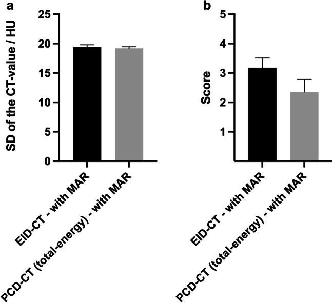
Quantitative and qualitative analysis demonstrated a lower degree of metal artifacts in the EID-CT images compared to the total-energy PCD-CT images (e.g., 82.99 ± 7.89 Hounsfield units (HU) versus 90.35 ± 6.28 HU; p < 0.001) with no qualitative difference between the high-energy bin PCD-CT images and the EID-CT images (4.18 ± 0.37 and 3.70 ± 0.64; p = 0.575). After MAR, artifacts were more profoundly reduced in the PCD-CT images compared to the EID-CT images in both analyses (e.g., 2.35 ± 0.43 and 3.18 ± 0.34; p < 0.001).

CONCLUSION

PCD-CT in combination with MAR have the potential to provide an improved option for reduction of coil-related artifacts in cerebral imaging in this in vitro study.

KEY POINTS

• Photon-counting detector CT produces more artifacts compared to energy-integrating detector CT without metal artifact reduction in cerebral in vitro imaging after neurovascular coil-embolization. • Spectral information of PCD-CT provides the potential for new post-processing techniques, since the coil-related artifacts were lower in PCD-CT images compared to EID-CT images after linear interpolation metal artifact reduction in this in vitro study.

摘要

目的

光子计数探测器 CT(PCD-CT)是 CT 成像的一种很有前途的新技术。本研究的目的是使用可比的标准脑成像协议,在金属伪影减少(MAR)前后,对 PCD-CT 和传统能量积分探测器 CT(EID-CT)的线圈相关伪影进行体外比较。

方法

将类似于脑动脉动脉瘤的橡胶乳胶结节填充有神经血管铂线圈,并插入脑成像体模中。使用标准脑成像协议,分别对 PCD-CT 和 EID-CT 进行了重复的图像采集和重建(分别为 10 次)。此外,对 PCD-CT 和 EID-CT 图像进行了线性插值 MAR。使用定量(圆环感兴趣区域的标准差)和定性(5 分制分析)方法分析了伪影的程度。

结果

定量和定性分析表明,EID-CT 图像的金属伪影程度低于总能量 PCD-CT 图像(例如,82.99±7.89HU 与 90.35±6.28HU;p<0.001),高能 bin PCD-CT 图像与 EID-CT 图像之间无定性差异(4.18±0.37 和 3.70±0.64;p=0.575)。MAR 后,在两种分析中,PCD-CT 图像中的伪影比 EID-CT 图像中的伪影减少得更为明显(例如,2.35±0.43 和 3.18±0.34;p<0.001)。

结论

在这项体外研究中,PCD-CT 结合 MAR 有可能为减少脑成像中的线圈相关伪影提供一种改进的选择。

关键要点

  • 在神经血管线圈栓塞后的体外脑成像中,PCD-CT 产生的伪影比 EID-CT 多,且未进行金属伪影减少处理。

  • 在这项体外研究中,经过线性插值金属伪影减少处理后,PCD-CT 图像中的线圈相关伪影低于 EID-CT 图像,这表明 PCD-CT 的光谱信息为新的后处理技术提供了潜力。

https://cdn.ncbi.nlm.nih.gov/pmc/blobs/7989/9889475/3e8de551252c/330_2022_9073_Fig5_HTML.jpg
https://cdn.ncbi.nlm.nih.gov/pmc/blobs/7989/9889475/e25df26ccf71/330_2022_9073_Fig1_HTML.jpg
https://cdn.ncbi.nlm.nih.gov/pmc/blobs/7989/9889475/cb6a30044c3e/330_2022_9073_Fig2_HTML.jpg
https://cdn.ncbi.nlm.nih.gov/pmc/blobs/7989/9889475/2632aa50fef2/330_2022_9073_Fig3_HTML.jpg
https://cdn.ncbi.nlm.nih.gov/pmc/blobs/7989/9889475/21a3f32bfbb6/330_2022_9073_Fig4_HTML.jpg
https://cdn.ncbi.nlm.nih.gov/pmc/blobs/7989/9889475/3e8de551252c/330_2022_9073_Fig5_HTML.jpg

相似文献

1
Metal artifacts and artifact reduction of neurovascular coils in photon-counting detector CT versus energy-integrating detector CT - in vitro comparison of a standard brain imaging protocol.光子计数探测器 CT 与能量积分探测器 CT 中神经血管线圈的金属伪影和伪影减少 - 标准脑成像协议的体外比较。
Eur Radiol. 2023 Feb;33(2):803-811. doi: 10.1007/s00330-022-09073-y. Epub 2022 Aug 20.
2
Reduction of Metal Artifacts and Improvement in Dose Efficiency Using Photon-Counting Detector Computed Tomography and Tin Filtration.使用基于单光子计数探测器的 CT 和锡滤过降低金属伪影并提高剂量效率。
Invest Radiol. 2019 Apr;54(4):204-211. doi: 10.1097/RLI.0000000000000535.
3
Quantifying lumen diameter in coronary artery stents with high-resolution photon counting detector CT and convolutional neural network denoising.利用高分辨率光子计数探测器 CT 和卷积神经网络降噪技术定量冠状动脉支架内腔直径。
Med Phys. 2023 Jul;50(7):4173-4181. doi: 10.1002/mp.16415. Epub 2023 Apr 17.
4
Multi-energy CT imaging for large patients using dual-source photon-counting detector CT.使用双源光子计数探测器 CT 对大患者进行多能量 CT 成像。
Phys Med Biol. 2020 Aug 31;65(17):17NT01. doi: 10.1088/1361-6560/ab99e4.
5
Photon-Counting Computed Tomography for Vascular Imaging of the Head and Neck: First In Vivo Human Results.基于单光子计数的头颈部血管 CT 成像:初步人体研究结果
Invest Radiol. 2018 Mar;53(3):135-142. doi: 10.1097/RLI.0000000000000418.
6
Metal artifact correction in photon-counting detector computed tomography: metal trace replacement using high-energy data.光子计数探测器计算机断层扫描中的金属伪影校正:使用高能数据进行金属痕迹替换
Med Phys. 2023 Jan;50(1):380-396. doi: 10.1002/mp.16049. Epub 2022 Oct 22.
7
Dose Reduction for Sinus and Temporal Bone Imaging Using Photon-Counting Detector CT With an Additional Tin Filter.使用带附加锡滤过器的光子计数探测器 CT 进行鼻窦和颞骨成像的剂量降低。
Invest Radiol. 2020 Feb;55(2):91-100. doi: 10.1097/RLI.0000000000000614.
8
Photon-Counting CT: High-Resolution Imaging of Coronary Stents.光子计数 CT:冠状动脉支架的高分辨率成像。
Invest Radiol. 2018 Mar;53(3):143-149. doi: 10.1097/RLI.0000000000000420.
9
CT Number Accuracy and Association With Object Size: A Phantom Study Comparing Energy-Integrating Detector CT and Deep Silicon Photon-Counting Detector CT.CT 数准确性与目标大小的关系:能量积分探测器 CT 与深硅光子计数探测器 CT 比较的体模研究。
AJR Am J Roentgenol. 2023 Oct;221(4):539-547. doi: 10.2214/AJR.23.29463. Epub 2023 May 31.
10
Photon-Counting Detector CT With Denoising for Imaging of the Osseous Pelvis at Low Radiation Doses: A Phantom Study.光子计数探测器 CT 联合降噪技术在低辐射剂量下骨盆骨成像中的应用:一项体模研究。
AJR Am J Roentgenol. 2024 Jan;222(1):e2329765. doi: 10.2214/AJR.23.29765. Epub 2023 Aug 30.

引用本文的文献

1
Photon-counting CT-angiography in comparison to digital subtraction angiography for assessing intracranial aneurysms after coiling or clipping.与数字减影血管造影术相比,光子计数CT血管造影术用于评估颅内动脉瘤栓塞或夹闭术后的情况。
Neuroradiology. 2025 Jun 2. doi: 10.1007/s00234-025-03650-w.
2
Investigation of radiology professionals' awareness of CT head artifacts.放射科专业人员对头部CT伪影的认知调查
BMC Res Notes. 2025 May 28;18(1):239. doi: 10.1186/s13104-025-07300-5.
3
In Vivo Discrimination of Iodine and Tantalum-Based Liquid Embolics After Intracranial or Spinal Embolization Using Photon-Counting Detector CT.
使用光子计数探测器CT对颅内或脊髓栓塞后基于碘和钽的液体栓塞剂进行体内鉴别
Clin Neuroradiol. 2025 Feb 6. doi: 10.1007/s00062-025-01502-x.
4
Photon-counting CT imaging of a patient with coiled and untreated intracranial saccular aneurysms.对一名患有盘绕状且未经治疗的颅内囊状动脉瘤患者进行的光子计数CT成像。
Neuroradiol J. 2025 Jan 29:19714009251313514. doi: 10.1177/19714009251313514.
5
Photon-counting computed tomography in radiology.放射学中的光子计数计算机断层扫描。
Pol J Radiol. 2024 Sep 11;89:e433-e442. doi: 10.5114/pjr/191743. eCollection 2024.
6
Dual Source Photon-Counting Computed Tomography-Part II: Clinical Overview of Neurovascular Applications.双源光子计数计算机断层扫描 - 第二部分:神经血管应用的临床概述
J Clin Med. 2023 May 23;12(11):3626. doi: 10.3390/jcm12113626.