CT R&D Group, Health & Medical Equipment Business, Samsung Electronics Co., Ltd., Suwon, Republic of Korea.
PLoS One. 2021 Mar 5;16(3):e0247355. doi: 10.1371/journal.pone.0247355. eCollection 2021.
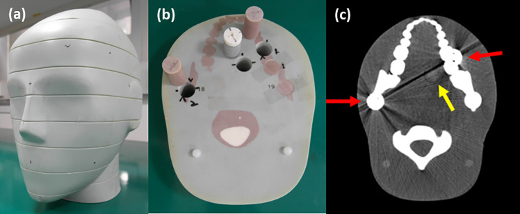
Metal artifacts are considered a major challenge in computed tomography (CT) as these adversely affect the diagnosis and treatment of patients. Several approaches have been developed to address this problem. The present study explored the clinical potential of a novel photon-counting detector (PCD) CT system in reducing metal artifacts in head CT scans. In particular, we studied the recovery of an oral tumor region located under metal artifacts after correction. Three energy thresholds were used to group data into three bins (bin 1: low-energy, bin 2: middle-energy, and bin 3: high-energy) in the prototype PCD CT system. Three types of physical phantoms were scanned on the prototype PCD CT system. First, we assessed the accuracy of iodine quantification using iodine phantoms at varying concentrations. Second, we evaluated the performance of material decomposition (MD) and virtual monochromatic images (VMIs) using a multi-energy CT phantom. Third, we designed an ATOM phantom with metal insertions to verify the effect of the proposed metal artifact reduction. In particular, we placed an insertion-mimicking an iodine-enhanced oral tumor in the beam path of metallic objects. Normalized metal artifact reduction (NMAR) was performed for each energy bin image, followed by an image-based MD and VMI reconstruction. Image quality was analyzed quantitatively by contrast-to-noise ratio (CNR) measurements. The results of iodine quantification showed a good match between the true and measured iodine concentrations. Furthermore, as expected, the contrast between iodine and the surrounding material was higher in bin 1 image than in bin 3 image. On the other hand, the bin 3 image of the ATOM phantom showed fewer metal artifacts than the bin 1 image because of the higher photon energy. The result of quantitative assessment demonstrated that the 40-keV VMI (CNR: 20.6 ± 1.2) with NMAR and MD remarkably increased the contrast of the iodine-enhanced region compared with that of the conventional images (CNR: 10.4 ± 0.5) having 30 to 140 keV energy levels. The PCD-based multi-energy CT imaging has immense potential to maximize the contrast of the target tissue and reduce metal artifacts simultaneously. We believe that it would open the door to novel applications for the diagnosis and treatment of several diseases.
金属伪影被认为是计算机断层扫描(CT)中的一个主要挑战,因为它们会对患者的诊断和治疗产生不利影响。已经开发了几种方法来解决这个问题。本研究探讨了新型光子计数探测器(PCD)CT 系统在减少头部 CT 扫描中金属伪影方面的临床潜力。特别是,我们研究了在校正后恢复位于金属伪影下的口腔肿瘤区域的能力。在原型 PCD CT 系统中,使用三个能量阈值将数据分为三个bins(bin 1:低能,bin 2:中能,bin 3:高能)。在原型 PCD CT 系统上扫描了三种物理体模。首先,我们使用不同浓度的碘体模评估碘定量的准确性。其次,我们使用多能 CT 体模评估材料分解(MD)和虚拟单色图像(VMI)的性能。第三,我们设计了一个带有金属插入物的 ATOM 体模,以验证所提出的减少金属伪影的效果。特别是,我们在金属物体的射束路径中放置了一个类似于碘增强口腔肿瘤的插入物。对每个能量 bin 图像进行归一化金属伪影减少(NMAR),然后进行基于图像的 MD 和 VMI 重建。通过对比噪声比(CNR)测量对图像质量进行定量分析。碘定量的结果表明,真实和测量的碘浓度之间有很好的匹配。此外,正如预期的那样,与周围材料相比,bin 1 图像中的碘对比度高于 bin 3 图像。另一方面,由于更高的光子能量,ATOM 体模的 bin 3 图像的金属伪影比 bin 1 图像少。定量评估的结果表明,与具有 30 至 140keV 能级的常规图像(CNR:10.4±0.5)相比,具有 40keV VMI(CNR:20.6±1.2)的 NMAR 和 MD 的图像显著增加了碘增强区域的对比度。基于 PCD 的多能 CT 成像具有很大的潜力,可以同时最大限度地提高目标组织的对比度并减少金属伪影。我们相信,它将为几种疾病的诊断和治疗开辟新的应用途径。