Abdelgalil Ahmed Ismael, Mohammed Faten Fathy
Department of Surgery, Anesthesiology and Radiology, Faculty of Veterinary Medicine, Cairo University, Giza, Egypt.
Department of Pathology, Faculty of Veterinary Medicine, Cairo University, Giza, Egypt.
Vet Res Forum. 2021;12(3):277-281. doi: 10.30466/vrf.2020.108341.2569. Epub 2021 Sep 15.
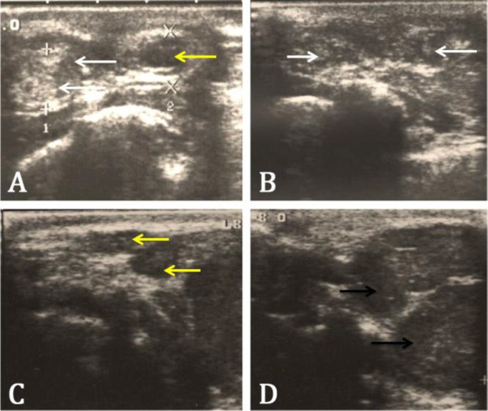
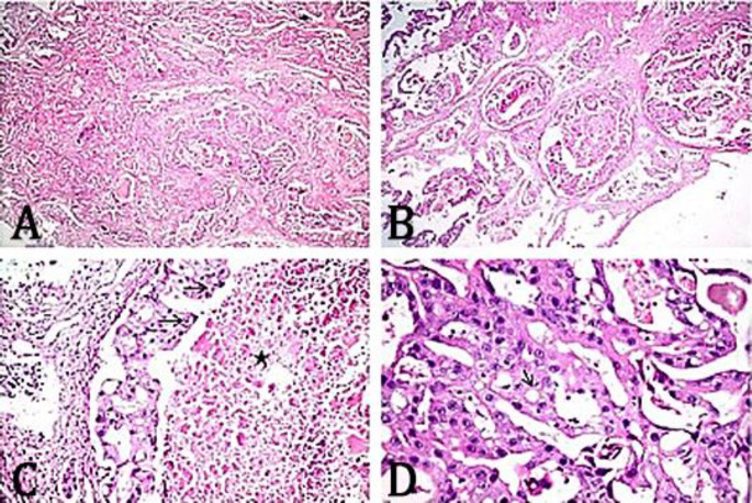
Ceruminous gland tumor is the most common tumor of the ear canal in cats. Otoscopic examination of the ear tumor is so difficult due to narrowing of the external ear canal. The present study aimed to investigate clinical, ultrasonographic and histopathological characteristics of feline ceruminous gland neoplasm in cats. Ten cats with unilateral ear canal swelling were subjected to thorough physical and clinical investigations. Ultrasound of the ear canal and parotid gland was performed using 8.00 MHz linear probe. Tissue specimens were collected after surgical excision (total ear canal ablation) for histopathological examination. Clinical examination of the ceruminous tumors revealed firm pinkish mass obliterated the ear canal with purulent or bloody aural discharge. Ultrasound examination of the ear tumor was helpful in detecting the size, shape, echogenicity and extension of the tumors to the surrounding structures as well as the nature of the feline ceruminous tumor. Histopathological examination was the main diagnostic tool for detecting the nature of the ceruminous neoplasms.
耵聍腺肿瘤是猫耳道最常见的肿瘤。由于外耳道狭窄,对耳部肿瘤进行耳镜检查非常困难。本研究旨在调查猫耵聍腺肿瘤的临床、超声和组织病理学特征。对10只单侧耳道肿胀的猫进行了全面的体格和临床检查。使用8.00 MHz线性探头对耳道和腮腺进行超声检查。手术切除(全耳道切除术)后收集组织标本进行组织病理学检查。耵聍肿瘤的临床检查显示,坚实的粉红色肿块阻塞了耳道,伴有脓性或血性耳漏。耳部肿瘤的超声检查有助于检测肿瘤的大小、形状、回声性以及肿瘤向周围结构的扩展情况,还有猫耵聍肿瘤的性质。组织病理学检查是检测耵聍肿瘤性质的主要诊断工具。