Siemens Medical Solutions USA, Inc., Knoxville, Tennessee, USA.
Advanced Clinical Imaging Technology, Siemens Healthcare AG, Lausanne, Switzerland.
Med Phys. 2022 Jan;49(1):309-323. doi: 10.1002/mp.15376. Epub 2021 Dec 10.
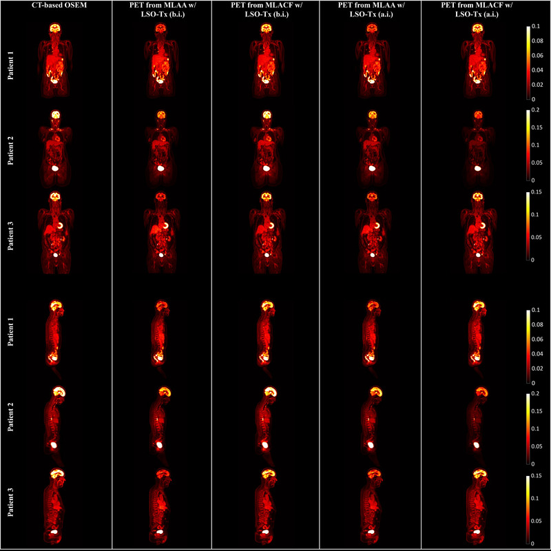
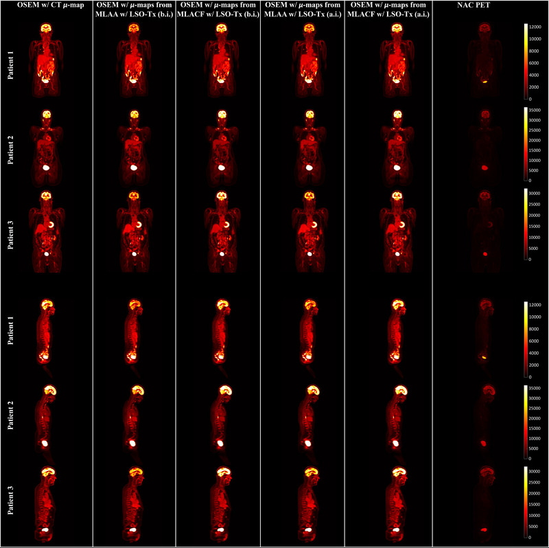
Long-axial field-of-view (FOV) positron emission tomography (PET) scanners have gained a lot of interest in the recent years. Such scanners provide increased sensitivity and enable unique imaging opportunities that were not previously feasible. Benefiting from the high sensitivity of a long-axial FOV PET scanner, we studied a computed tomography (CT)-less reconstruction algorithm for the Siemens Biograph Vision Quadra with an axial FOV of 106 cm.
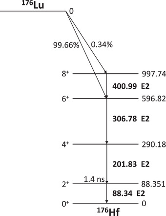
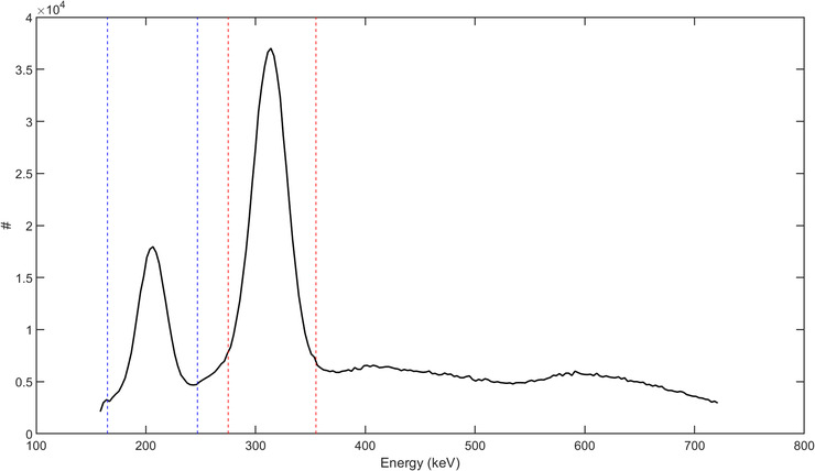
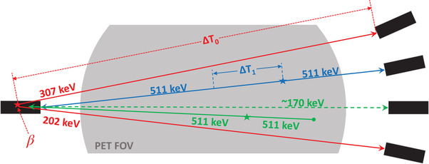
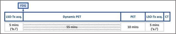
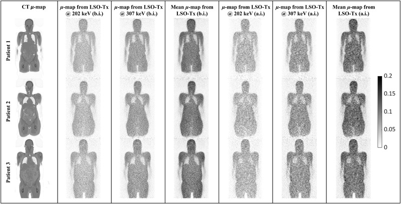
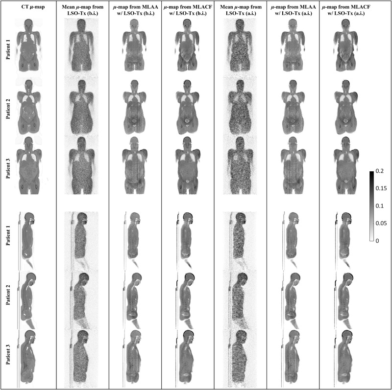
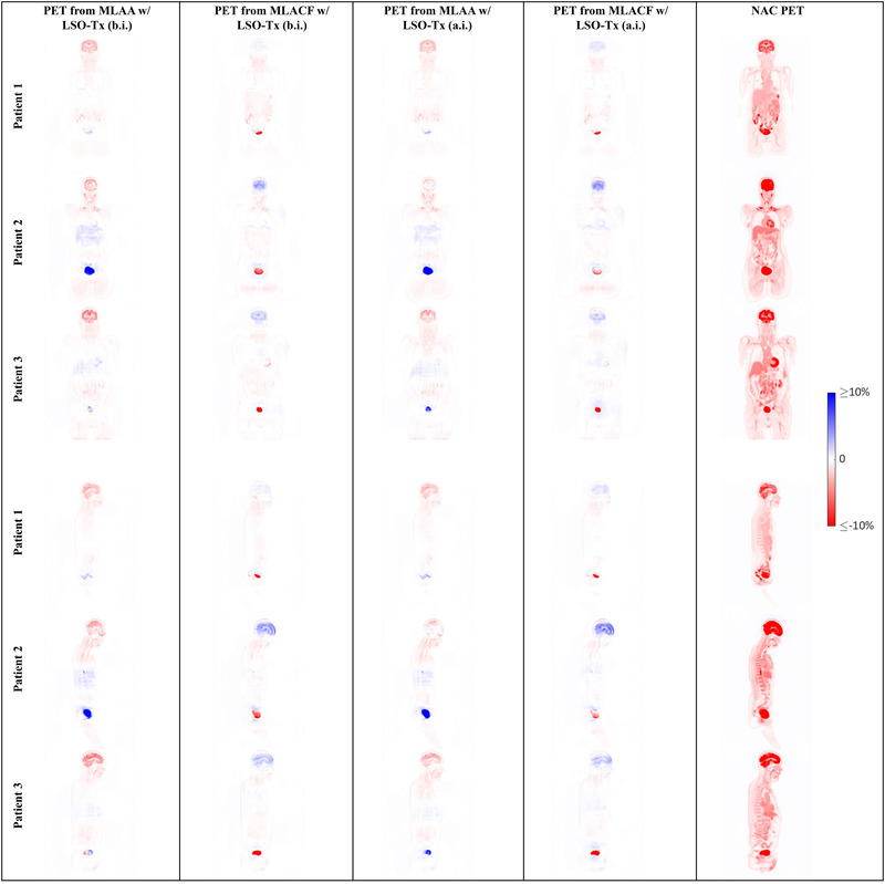
In this work, the background radiation from radioisotope lutetium-176 in the scintillators was used to create an initial estimate of the attenuation maps. Then, joint activity and attenuation reconstruction algorithms were used to create an improved attenuation map of the object. The final attenuation maps were then used to reconstruct quantitative PET images, which were compared against CT-based PET images. The proposed method was evaluated on data from three patients who underwent a flurodeoxyglucouse PET scan.
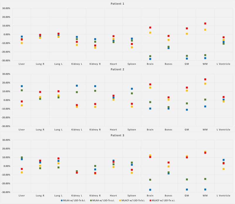
Segmentation of the PET images of the three studied patients showed an average quantitative error of 6.5%-8.3% across all studied organs when using attenuation maps from maximum likelihood estimation of attenuation and activity and 5.3%-6.6% when using attenuation maps from maximum likelihood estimation of activity and attenuation correction coefficients.
Benefiting from the background radiation of lutetium-based scintillators, a quantitative CT-less PET imaging technique was evaluated in this work.
近年来,长轴向视野(FOV)正电子发射断层扫描(PET)扫描仪引起了广泛关注。此类扫描仪提高了灵敏度,并提供了以前无法实现的独特成像机会。得益于长轴向 FOV PET 扫描仪的高灵敏度,我们研究了一种适用于轴向 FOV 为 106cm 的西门子 Biograph Vision Quadra 的无 CT 重建算法。
在这项工作中,使用来自闪烁体中的放射性同位素镥-176 的本底辐射来创建衰减图的初始估计。然后,使用联合活动和衰减重建算法来创建物体的改进衰减图。最后,使用最终的衰减图来重建定量 PET 图像,并与基于 CT 的 PET 图像进行比较。该方法在 3 名接受氟脱氧葡萄糖 PET 扫描的患者的数据上进行了评估。
对 3 名研究患者的 PET 图像进行分割,当使用最大似然估计衰减和活动的衰减图时,所有研究器官的平均定量误差为 6.5%-8.3%,当使用最大似然估计活动和衰减校正系数的衰减图时,平均定量误差为 5.3%-6.6%。
本研究评估了一种基于镥基闪烁体本底辐射的定量无 CT PET 成像技术。