DeFroda Steven F, Alter Thomas D, Lambers Floor, Malloy Philip, Clapp Ian M, Chahla Jorge, Nho Shane J
Section of Young Adult Hip Surgery, Division of Sports Medicine, Department of Orthopaedic Surgery, Hip Preservation Center, Rush University Medical Center, Chicago, Illinois, USA.
Stryker Sports Medicine, Kalamazoo, Michigan, USA.
Orthop J Sports Med. 2021 Nov 19;9(11):23259671211049457. doi: 10.1177/23259671211049457. eCollection 2021 Nov.
Accurate assessment of osseous morphology is imperative in the evaluation of patients with femoroacetabular impingement syndrome (FAIS) and hip dysplasia. Through use of computed tomography (CT), 3-dimensional (3D) reconstructed hip models may provide a more precise measurement for overcoverage and undercoverage and aid in the interpretation of 2-dimensional radiographs obtained in the clinical setting.
To describe new measures of acetabular coverage based on 3D-reconstructed CT scan bone models.
Cross-sectional study; Level of evidence, 3.
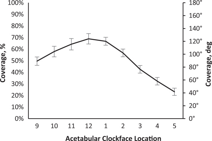
Preoperative CT scans were acquired on the bilateral hips and pelvises of 30 patients before arthroscopic surgical intervention for FAIS. Custom software was used for semiautomated segmentation to generate 3D osseous models of the femur and acetabulum that were aligned to a standard coordinate system. This software calculated percentage of total acetabular coverage, which was defined as the surface area projected onto the superior aspect of the femoral head. The percentage of coverage was also quantified regionally in the anteromedial, anterolateral, posteromedial, and posterolateral quadrants of the femoral head. The acetabular clockface was established by defining 6 o'clock as the inferior aspect of the acetabular notch. Radial coverage was then calculated along the clockface from the 9-o'clock to 5-o'clock positions.
The study included 20 female and 10 male patients with a mean age of 33.6 ± 11.7 years and mean body mass index of 27.8 ± 6.3. The average percentage of total acetabular coverage for the sample was 57% ± 6%. Acetabular coverages by region were as follows: anteromedial, 78% ± 7%; anterolateral, 18% ± 7%, posterolateral, 33% ± 13%, and posteromedial, 99% ± 1%. The acetabular coverage ranged from 23% to 69% along the radial clockface from 9 to 5 o'clock.
This study demonstrated new 3D measurements to characterize acetabular coverage in patients with FAIS and elucidated the distribution of acetabular coverage according to these measurements.
在评估股骨髋臼撞击综合征(FAIS)和髋关节发育不良患者时,准确评估骨形态至关重要。通过使用计算机断层扫描(CT),三维(3D)重建的髋关节模型可以为髋臼覆盖率过高和过低提供更精确的测量,并有助于解释临床环境中获得的二维X线片。
描述基于3D重建CT扫描骨模型的髋臼覆盖率新测量方法。
横断面研究;证据等级,3级。
对30例因FAIS接受关节镜手术干预的患者,在术前对其双侧髋关节和骨盆进行CT扫描。使用定制软件进行半自动分割,以生成与标准坐标系对齐的股骨和髋臼的3D骨模型。该软件计算髋臼总覆盖率,定义为投影到股骨头上方的表面积。覆盖率还在股骨头的前内侧、前外侧、后外侧和后内侧象限进行区域量化。通过将髋臼切迹的下侧定义为6点来建立髋臼钟面。然后从9点到5点沿钟面计算径向覆盖率。
该研究纳入了20名女性和10名男性患者,平均年龄为33.6±11.7岁,平均体重指数为27.8±6.3。样本的髋臼总覆盖率平均为57%±6%。各区域的髋臼覆盖率如下:前内侧,78%±7%;前外侧,18%±7%,后外侧,33%±13%,后内侧,99%±1%。从9点到5点沿径向钟面的髋臼覆盖率范围为23%至69%。
本研究展示了用于表征FAIS患者髋臼覆盖率的新3D测量方法,并根据这些测量结果阐明了髋臼覆盖率的分布情况。