Department of Radiology, Johns Hopkins University School of Medicine, Baltimore, MD, USA.
Department of Neurology, Johns Hopkins University School of Medicine, Baltimore, MD, USA.
Neuroimage. 2021 Dec 15;245:118754. doi: 10.1016/j.neuroimage.2021.118754. Epub 2021 Nov 23.
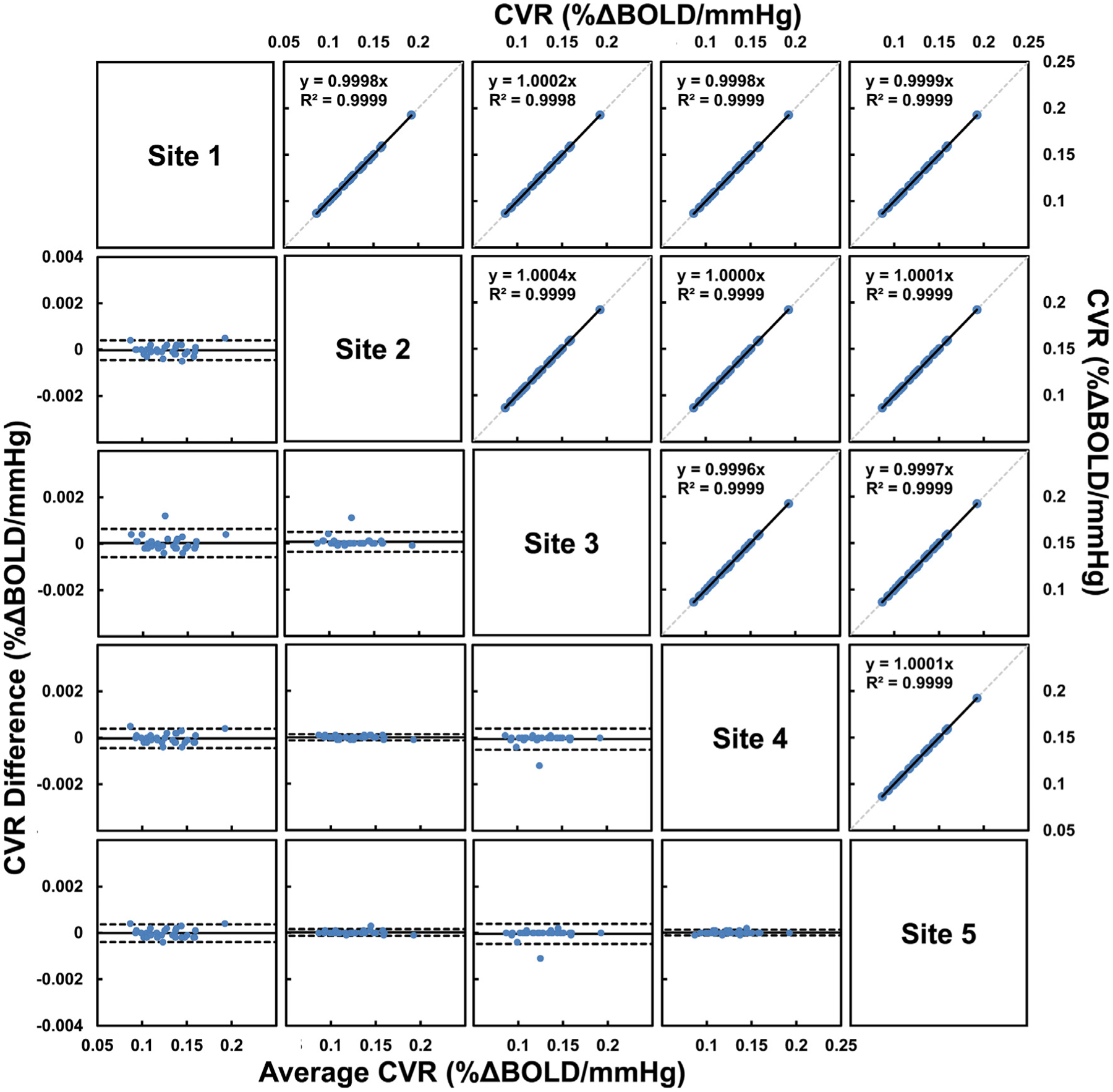
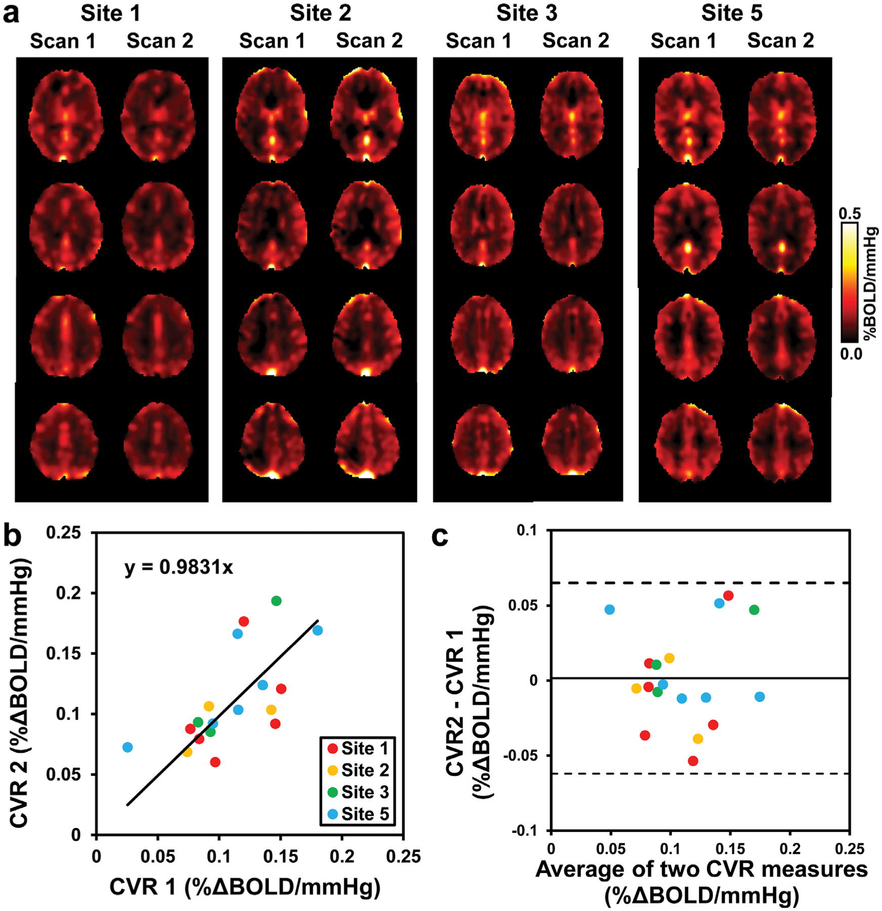
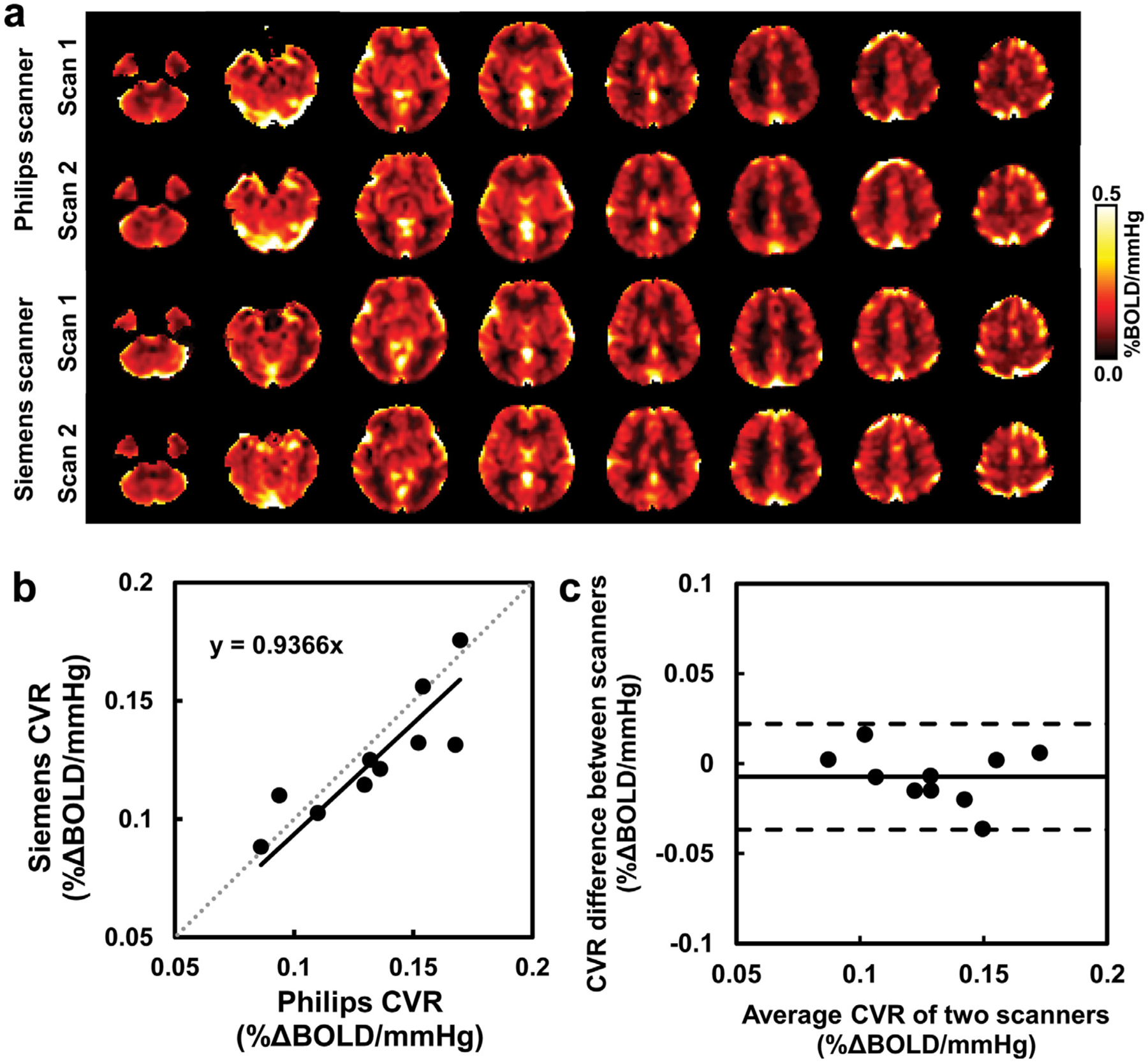
Cerebrovascular reactivity (CVR), which measures the ability of cerebral blood vessels to dilate or constrict in response to vasoactive stimuli such as CO2 inhalation, is an important index of the brain's vascular health. Quantification of CVR using BOLD MRI with hypercapnia challenge has shown great promises in research and clinical studies. However, in order for it to be used as a potential imaging biomarker in large-scale and multi-site studies, the reliability of CO2-CVR quantification across different MRI acquisition platforms and researchers/raters must be examined. The goal of this report from the MarkVCID small vessel disease biomarkers consortium is to evaluate the reliability of CO2-CVR quantification in three studies. First, the inter-rater reliability of CO2-CVR data processing was evaluated by having raters from 5 MarkVCID sites process the same 30 CVR datasets using a cloud-based CVR data processing pipeline. Second, the inter-scanner reproducibility of CO2-CVR quantification was assessed in 10 young subjects across two scanners of different vendors. Third, test-retest repeatability was evaluated in 20 elderly subjects from 4 sites with a scan interval of less than 2 weeks. In all studies, the CO2 CVR measurements were performed using the fixed inspiration method, where the subjects wore a nose clip and a mouthpiece and breathed room air and 5% CO2 air contained in a Douglas bag alternatively through their mouth. The results showed that the inter-rater CoV of CVR processing was 0.08 ± 0.08% for whole-brain CVR values and ranged from 0.16% to 0.88% in major brain regions, with ICC of absolute agreement above 0.9959 for all brain regions. Inter-scanner CoV was found to be 6.90 ± 5.08% for whole-brain CVR values, and ranged from 4.69% to 12.71% in major brain regions, which are comparable to intra-session CoVs obtained from the same scanners on the same day. ICC of consistency between the two scanners was 0.8498 for whole-brain CVR and ranged from 0.8052 to 0.9185 across major brain regions. In the test-retest evaluation, test-retest CoV across different days was found to be 18.29 ± 17.12% for whole-brain CVR values, and ranged from 16.58% to 19.52% in major brain regions, with ICC of absolute agreement ranged from 0.6480 to 0.7785. These results demonstrated good inter-rater, inter-scanner, and test-retest reliability in healthy volunteers, and suggested that CO2-CVR has suitable instrumental properties for use as an imaging biomarker of cerebrovascular function in multi-site and longitudinal observational studies and clinical trials.
脑血管反应性(CVR)是衡量脑血管在对血管活性刺激(如 CO2 吸入)反应时扩张或收缩能力的重要指标,是大脑血管健康的一个重要指标。使用 BOLD MRI 进行二氧化碳挑战来定量 CVR 在研究和临床研究中显示出了很大的前景。然而,为了使其能够在大规模和多站点研究中作为潜在的成像生物标志物使用,必须检验不同 MRI 采集平台和研究人员/评分者之间 CO2-CVR 定量的可靠性。该报告来自 MarkVCID 小血管疾病生物标志物联盟的目标是评估三个研究中 CO2-CVR 定量的可靠性。首先,通过让来自 5 个 MarkVCID 站点的评分者使用基于云的 CVR 数据处理管道处理相同的 30 个 CVR 数据集,评估了 CO2-CVR 数据处理的评分者间可靠性。其次,在两台不同供应商的扫描仪上评估了 10 名年轻受试者的 CO2-CVR 定量的扫描仪间可重复性。最后,在 4 个站点的 20 名老年受试者中评估了测试-重测的可重复性,扫描间隔小于 2 周。在所有研究中,使用固定吸气方法进行 CO2 CVR 测量,受试者佩戴鼻夹和口器,通过口交替呼吸室内空气和 5% CO2 空气袋中的空气。结果表明,CVR 处理的评分者间 CoV 为全脑 CVR 值的 0.08±0.08%,主要脑区的范围为 0.16%至 0.88%,所有脑区的绝对一致性 ICC 均高于 0.9959。扫描仪间 CoV 为全脑 CVR 值的 6.90±5.08%,主要脑区的范围为 4.69%至 12.71%,与同一天在同一扫描仪上获得的单次会话 CoV 相当。两台扫描仪之间的一致性 ICC 为全脑 CVR 的 0.8498,主要脑区的范围为 0.8052 至 0.9185。在测试-重测评估中,不同日期的测试-重测 CoV 发现全脑 CVR 值为 18.29±17.12%,主要脑区的范围为 16.58%至 19.52%,绝对一致性 ICC 范围为 0.6480 至 0.7785。这些结果表明在健康志愿者中具有良好的评分者间、扫描仪间和测试-重测可靠性,提示 CO2-CVR 具有适合作为多站点和纵向观察性研究和临床试验中脑血管功能成像生物标志物的仪器特性。