Centre Universitaire d'Ophtalmologie-Recherche (CUO-Recherche), Centre de Recherche du CHU de Québec, Axe Médecine Régénératrice, Hôpital du Saint-Sacrement, Québec, QC G1S 4L8, Canada.
Centre de Recherche en Organogénèse Expérimentale de l'Université Laval/LOEX, Génie Tissulaire et Régénération, Centre de Recherche du CHU de Québec, Axe Médecine Régénératrice, Québec, QC G1V 0A6, Canada.
Int J Mol Sci. 2021 Nov 17;22(22):12426. doi: 10.3390/ijms222212426.
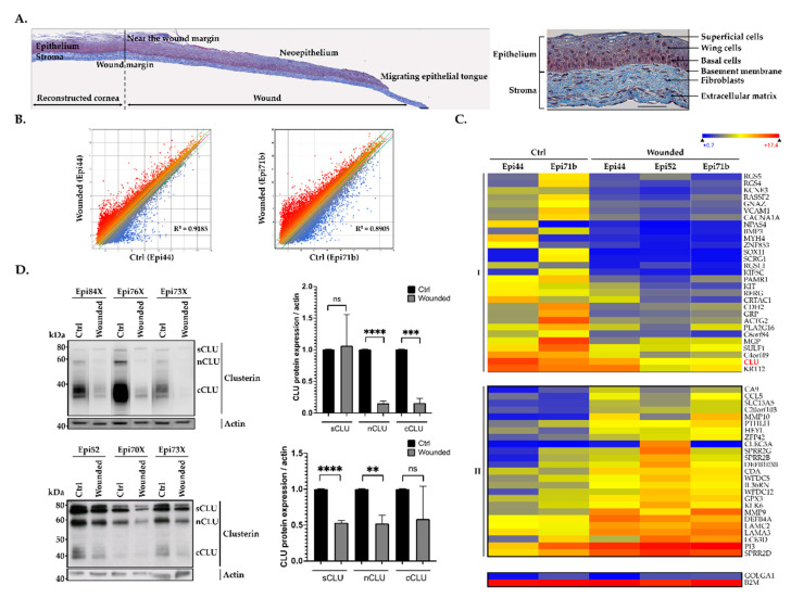
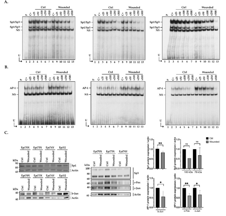
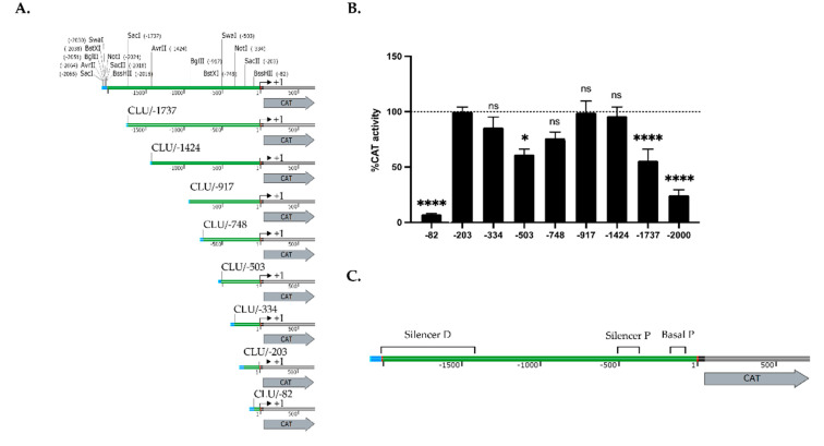
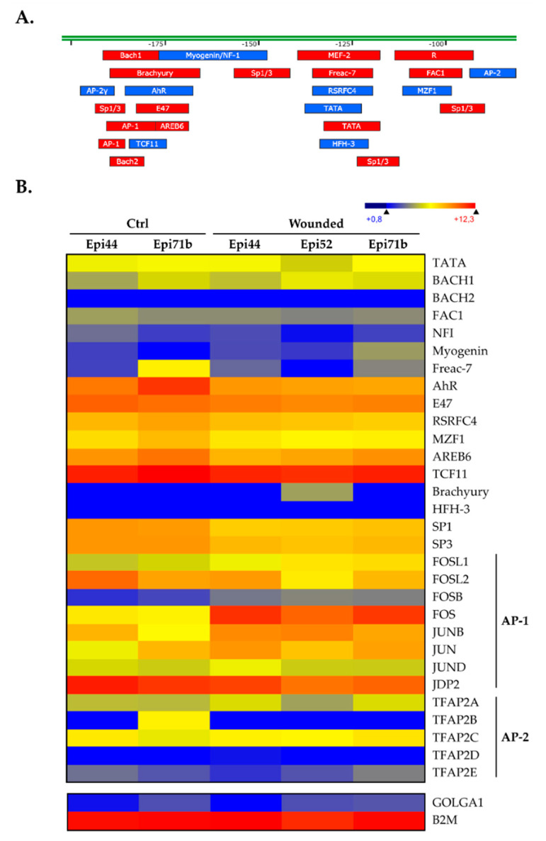
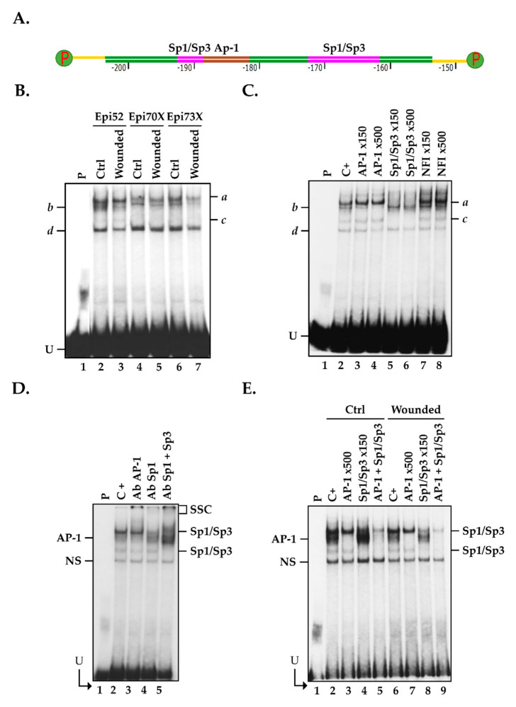
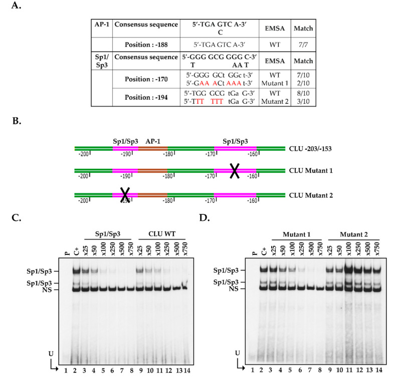
In order to reduce the need for donor corneas, understanding of corneal wound healing and development of an entirely tissue-engineered human cornea (hTECs) is of prime importance. In this study, we exploited the hTEC to determine how deep wound healing affects the transcriptional pattern of corneal epithelial cells through microarray analyses. We demonstrated that the gene encoding () has its expression dramatically repressed during closure of hTEC wounds. Western blot analyses confirmed a strong reduction in the expression of the isoforms after corneal damage and suggest that repression of gene expression might be a prerequisite to hTEC wound closure. Transfection with segments from the human gene promoter revealed the presence of three regulatory regions: a basal promoter and two more distal negative regulatory regions. The basal promoter bears DNA binding sites for very potent transcription factors (TFs): Activator Protein-1 (AP-1) and Specificity protein-1 and 3 (Sp1/Sp3). By exploiting electrophoretic mobility shift assays (EMSA), we demonstrated that AP-1 and Sp1/Sp3 have their DNA binding site overlapping with one another in the basal promoter of the gene in hCECs. Interestingly, expression of both these TFs is reduced (at the protein level) during hTEC wound healing, thereby contributing to the extinction of gene expression during that process. The results of this study contribute to a better understanding of the molecular mechanisms accounting for the repression of gene expression during corneal wound healing.
为了减少对供体角膜的需求,理解角膜伤口愈合和开发完全组织工程化的人角膜(hTEC)至关重要。在这项研究中,我们利用 hTEC 来确定深度伤口愈合如何通过微阵列分析影响角膜上皮细胞的转录模式。我们表明,编码 () 的基因在 hTEC 伤口闭合过程中其表达被显著抑制。Western blot 分析证实,角膜损伤后 同工型的表达强烈减少,并表明 基因表达的抑制可能是人 TEC 伤口闭合的前提。用来自人 基因启动子的片段转染显示存在三个调节区:基本启动子和两个更远端的负调节区。基本启动子具有与非常有效的转录因子(TFs)结合的 DNA 结合位点:激活蛋白-1 (AP-1) 和特异性蛋白-1 和 3 (Sp1/Sp3)。通过利用电泳迁移率变动分析 (EMSA),我们证明在 hCEC 中的 基因的基本启动子中,AP-1 和 Sp1/Sp3 的 DNA 结合位点彼此重叠。有趣的是,这两种 TF 的表达在 hTEC 伤口愈合过程中(在蛋白质水平上)降低,从而有助于在该过程中 基因表达的消失。这项研究的结果有助于更好地理解在角膜伤口愈合过程中抑制 基因表达的分子机制。