Beya M F, Miyasaka M, Dudler L, Ezaki T, Trnka Z
Immunology. 1986 Jan;57(1):115-21.
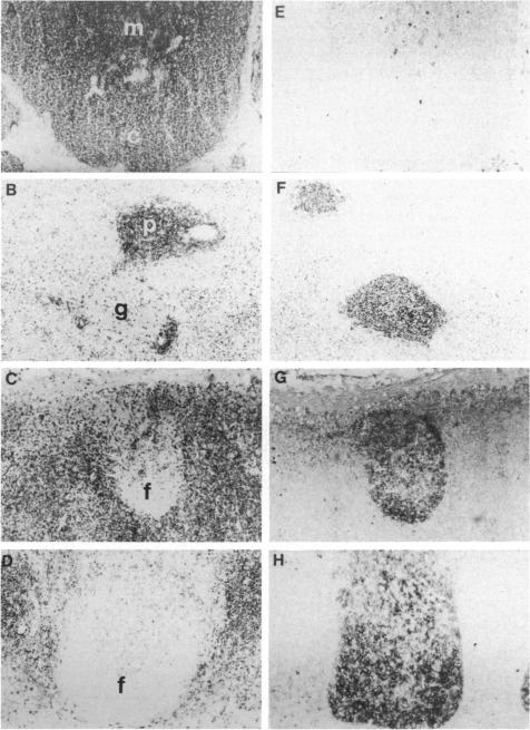
Two mouse monoclonal cytotoxic antibodies (ST-1a and ST-1b) recognize an antigen present on the large majority of thymocytes and all T cells in the periphery, but not B cells or other haemopoietic cells in sheep. Examination of frozen sections of various fetal tissues revealed that the cells expressing this antigen first appeared in the thymus, and these cells markedly increase in numbers in the peripheral lymphoid tissues after mid-gestation. Large accumulations of positive cells were located in the paracortex of lymph nodes, the periarteriolar lymphoid sheath of the spleen, and interfollicular areas of jejunal Peyer's patches, all of which are known to be T-dependent areas. Treatment of lymphocytes with ST-1a and complement resulted in the abrogation of T-proliferative responses, but the response to a B-cell mitogen, lipopolysaccharide, was not reduced. Neither ST-1a nor ST-1b cross-reacted to lymphocytes obtained from other species of animals (man, monkey, mouse, rat, guinea-pig, chicken, frog, pig, horse, goat and cattle). Based on these findings, it was concluded that the expression of the antigen recognized by ST-1a and ST-1b is restricted to the T-cell lineage of sheep, and that all ovine T cells express this antigen. Furthermore, ST-1a and ST-1b were determined to recognize the same antigen by reciprocal blocking experiments.
两种小鼠单克隆细胞毒性抗体(ST-1a和ST-1b)识别一种存在于绝大多数胸腺细胞以及外周所有T细胞上的抗原,但不识别绵羊的B细胞或其他造血细胞。对各种胎儿组织的冰冻切片检查显示,表达这种抗原的细胞首先出现在胸腺中,并且在妊娠中期后这些细胞在外周淋巴组织中的数量显著增加。大量阳性细胞聚集在淋巴结的副皮质区、脾脏的动脉周围淋巴鞘以及空肠派伊尔结的滤泡间区,所有这些区域都是已知的T细胞依赖区。用ST-1a和补体处理淋巴细胞导致T细胞增殖反应被消除,但对B细胞有丝分裂原脂多糖的反应并未降低。ST-1a和ST-1b都不与从其他动物物种(人、猴、小鼠、大鼠、豚鼠、鸡、青蛙、猪、马、山羊和牛)获得的淋巴细胞发生交叉反应。基于这些发现,得出结论:ST-1a和ST-1b识别的抗原表达仅限于绵羊的T细胞谱系,并且所有绵羊T细胞都表达这种抗原。此外,通过相互阻断实验确定ST-1a和ST-1b识别相同的抗原。