Department of Periodontics and Oral Medicine, University of Michigan, Ann Arbor, Michigan, USA.
University of Michigan Rogel Cancer Center, Ann Arbor, Michigan, USA.
Oncoimmunology. 2021 Nov 13;10(1):1997385. doi: 10.1080/2162402X.2021.1997385. eCollection 2021.
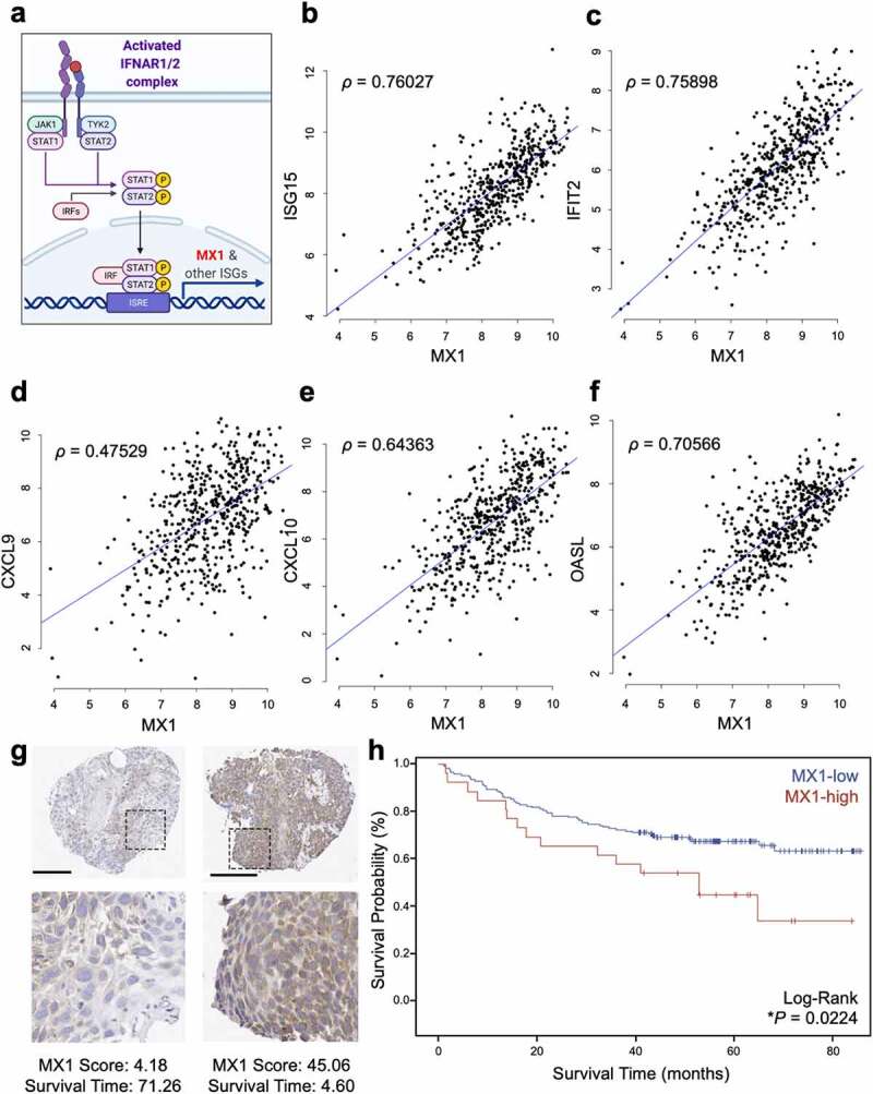
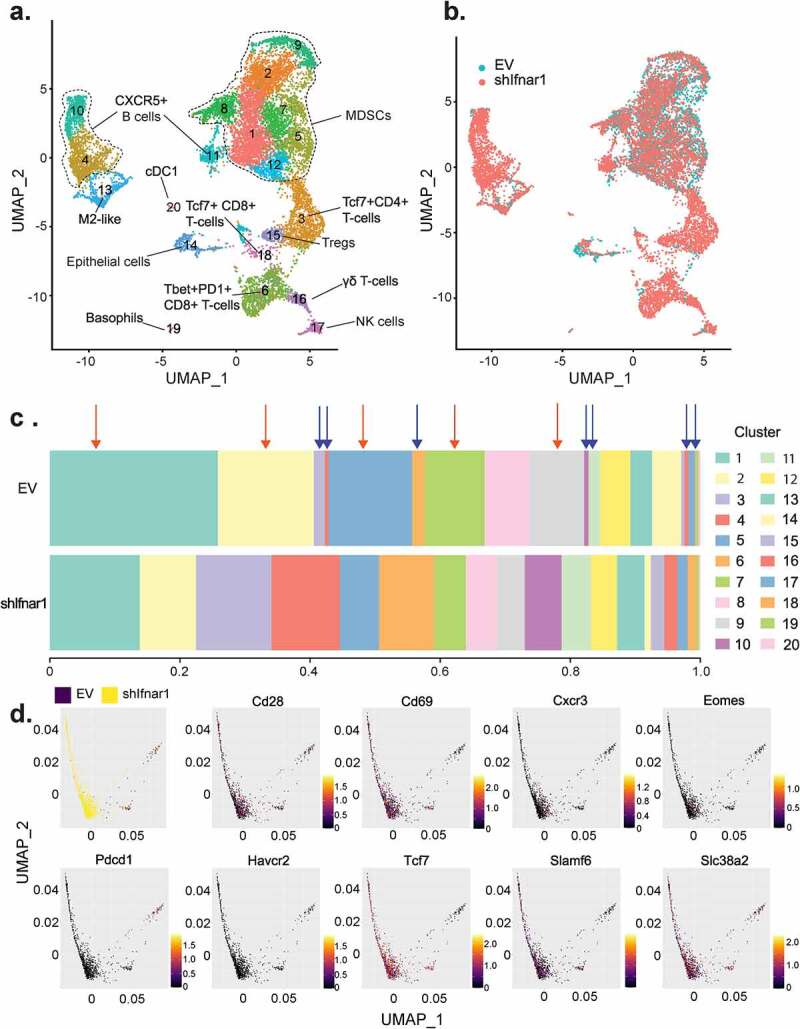
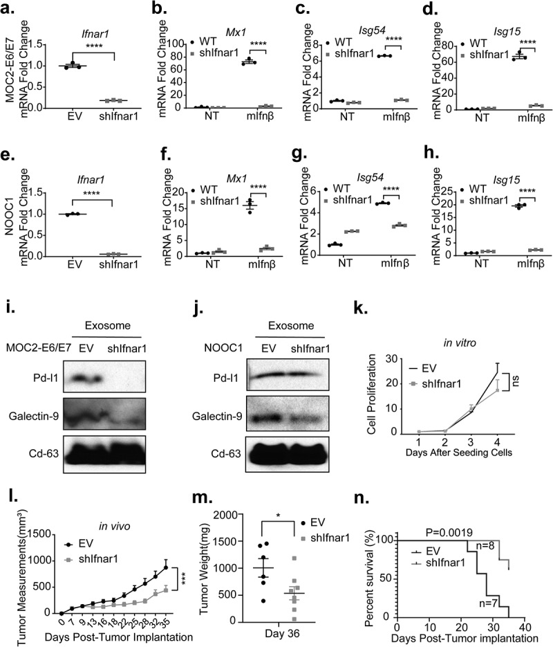
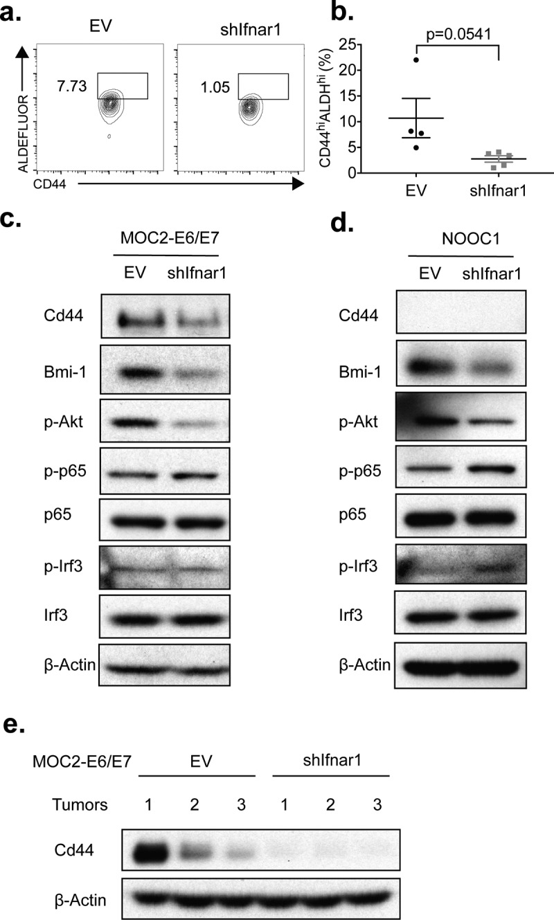
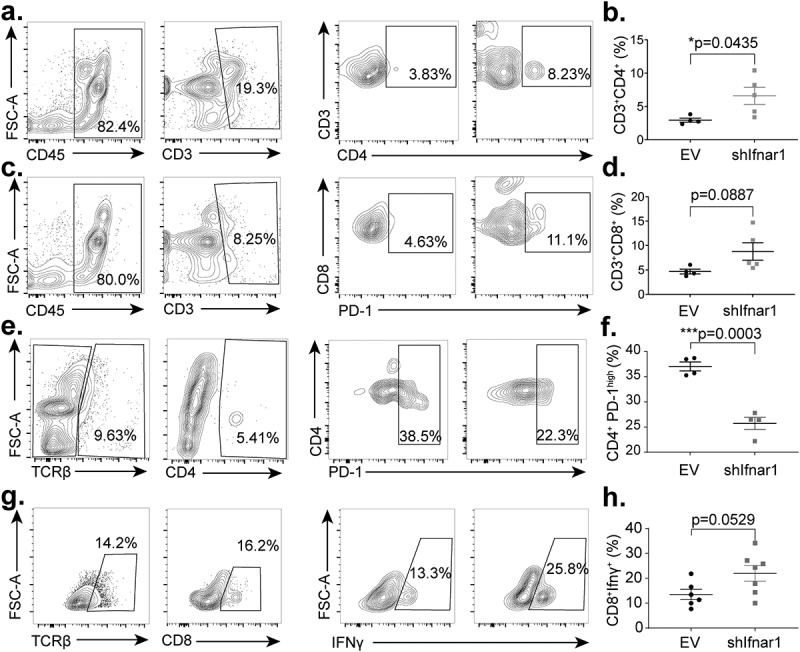
Type-I interferon (IFN-I) signaling is critical to maintaining antigen-presenting cell function for anti-tumor immunity. However, recent studies have suggested that IFN-I signaling may also contribute to more aggressive phenotypes, raising the possibility that IFN-I downstream signaling in cancer and myeloid cells may exert dichotomous functions.We analyzed the clinicopathologic correlation of cancer-specific IFN-I activation in 195 head and neck squamous cell carcinoma patients. We also characterized the immune impact of IFN-I receptor (IFNAR1)-deficiency in syngeneic tumor models using biochemistry, flow cytometry, and single-cell RNA-Seq. We stained HNSCC tissue microarrays with a sensitive IFN-I downstream signaling activation marker, MX1, and quantitated cancer cell-specific MX1 staining. Kaplan-Meier analysis revealed that MX1-high tumors exhibited worse survival, a phenotype that depends on the number of CD8 intratumoral T-cells. We found that cancer-specific IFNAR1 engagement promotes cancer stemness and higher expression levels of suppressive immune checkpoint receptor ligands in cancer-derived exosomes. Notably, mice bearing -deficient tumors exhibited lower tumor burden, increased T-cell infiltration, reduced exhausted CD4PD1 T-cells, and increased effector population CD8IFN-γ T-cells. Then, we performed single-cell RNA-sequencing and discovered that cancer-specific IFN-I signaling not only restricts effector cells expansion but also dampens their functional fitness.The beneficial role of IFN-I activation is largely dependent on the myeloid compartment. Cancer-specific IFN-I receptor engagement promotes cancer stemness and the release of cancer-derived exosomes with high expression levels of immune checkpoint receptor ligands. Cancer-specific IFN-I activation is associated with poor immunogenicity and worse clinical outcomes in HNSCC.
I 型干扰素 (IFN-I) 信号对于维持抗原呈递细胞的抗肿瘤免疫功能至关重要。然而,最近的研究表明,IFN-I 信号也可能导致更具侵袭性的表型,这提示癌症和髓系细胞中的 IFN-I 下游信号可能发挥双重作用。我们分析了 195 例头颈部鳞状细胞癌患者中癌症特异性 IFN-I 激活的临床病理相关性。我们还使用生物化学、流式细胞术和单细胞 RNA-Seq 对同种异体肿瘤模型中 IFN-I 受体 (IFNAR1) 缺陷的免疫影响进行了表征。我们使用一种敏感的 IFN-I 下游信号激活标记物 MX1 对 HNSCC 组织微阵列进行染色,并定量分析了癌症细胞特异性 MX1 染色。Kaplan-Meier 分析显示,MX1 高肿瘤的生存预后较差,这种表型取决于肿瘤内 CD8 T 细胞的数量。我们发现,癌症特异性 IFNAR1 结合促进了癌症干性和癌症衍生外泌体中更高水平的抑制性免疫检查点受体配体的表达。值得注意的是,携带 -缺陷肿瘤的小鼠表现出更低的肿瘤负担、增加的 T 细胞浸润、减少的耗尽 CD4PD1 T 细胞和增加的效应细胞群体 CD8IFN-γ T 细胞。然后,我们进行了单细胞 RNA-seq 测序,发现癌症特异性 IFN-I 信号不仅限制了效应细胞的扩增,还抑制了它们的功能适应性。IFN-I 激活的有益作用在很大程度上取决于髓系细胞。癌症特异性 IFNAR1 结合促进了癌症干性和具有高免疫检查点受体配体表达水平的癌症衍生外泌体的释放。癌症特异性 IFN-I 激活与 HNSCC 中的低免疫原性和较差的临床结果相关。