Department of Otorhinolaryngology, University Hospital Essen, University Duisburg-Essen, Essen, Germany.
Institute for Experimental Immunology and Imaging, University Duisburg-Essen, Essen, Germany.
Front Immunol. 2022 Jun 27;13:878959. doi: 10.3389/fimmu.2022.878959. eCollection 2022.
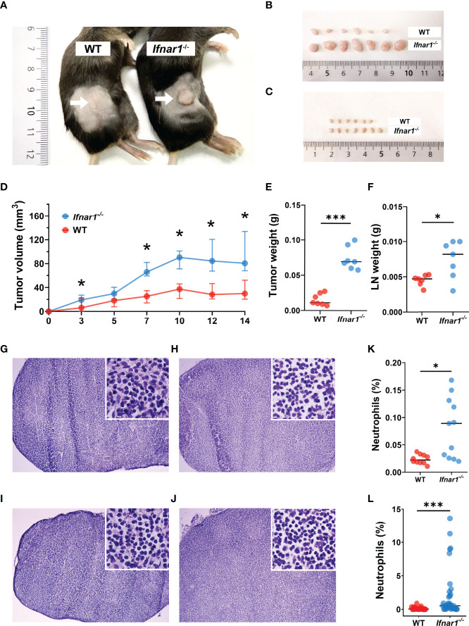
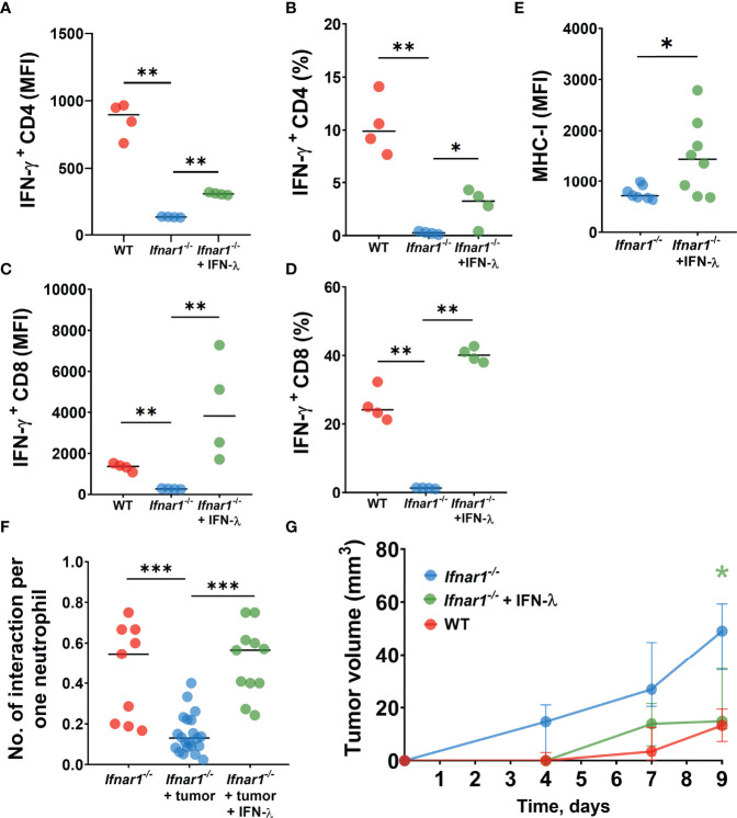
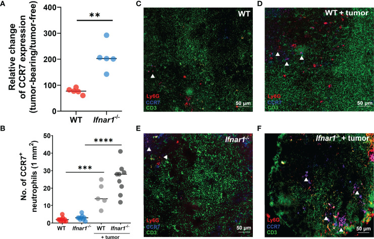
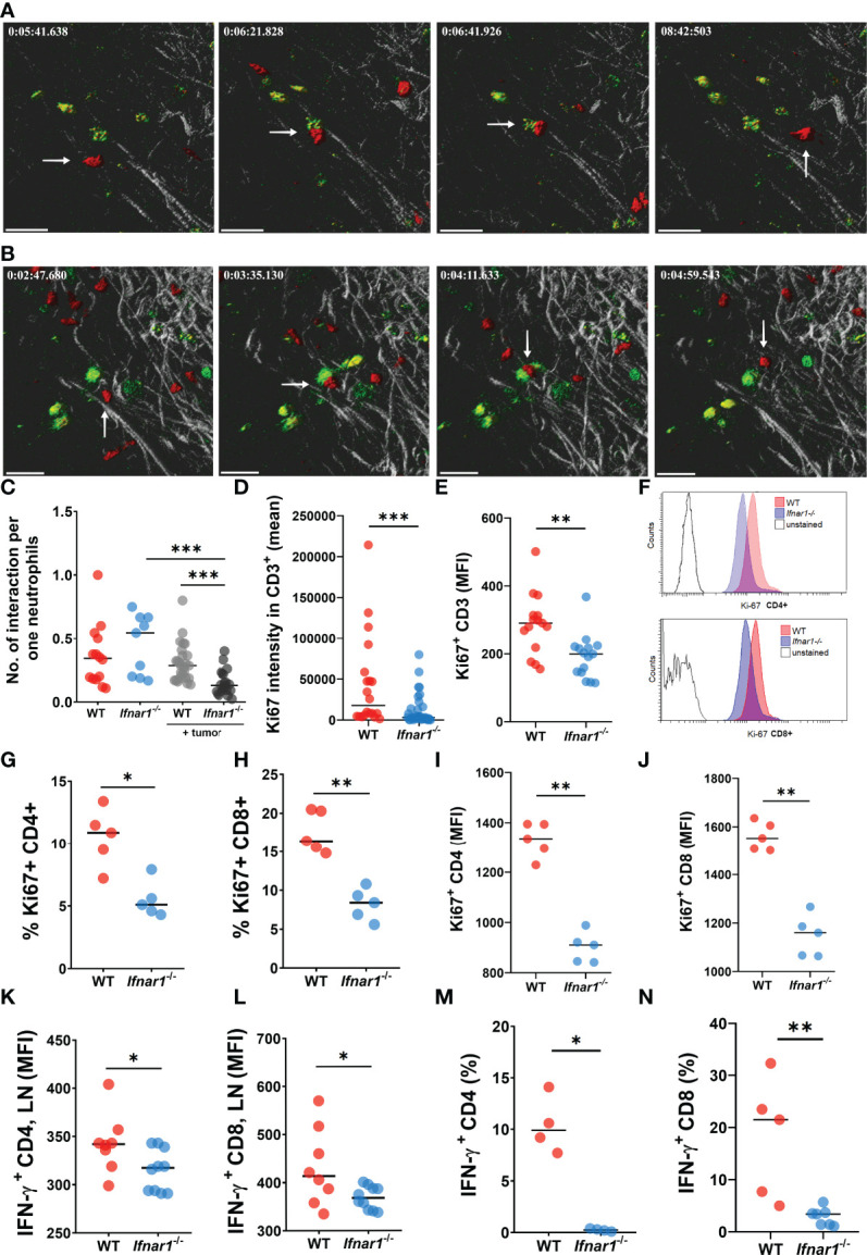
Tumor-draining lymph nodes (TDLNs) are the first organs where the metastatic spread of different types of cancer, including head and neck cancer (HNC), occurs and have therefore high prognostic relevance. Moreover, first anti-cancer immune responses have been shown to be initiated in such LNs tumor-educated myeloid cells. Among myeloid cells present in TDLNs, neutrophils represent a valuable population and considerably participate in the activation of effector lymphocytes there. Tumor-supportive or tumor-inhibiting activity of neutrophils strongly depends on the surrounding microenvironment. Thus, type I interferon (IFN) availability has been shown to prime anti-tumor activity of these cells. In accordance, mice deficient in type I IFNs show elevated tumor growth and metastatic spread, accompanied by the pro-tumoral neutrophil bias. To reveal the mechanism responsible for this phenomenon, we have studied here the influence of defective type I IFN signaling on the immunoregulatory activity of neutrophils in TDLNs. Live imaging of such LNs was performed using two-photon microscopy in a transplantable murine HNC model. Catchup and (type I IFN receptor- deficient) Catchup mice were used to visualize neutrophils and to assess their interaction with T-cells . We have evaluated spatiotemporal patterns of neutrophil/T-cell interactions in LNs in the context of type I interferon receptor (IFNAR1) availability in tumor-free and tumor-bearing animals. Moreover, phenotypic and functional analyses were performed to further characterize the mechanisms regulating neutrophil immunoregulatory capacity. We demonstrated that inactive IFNAR1 leads to elevated accumulation of neutrophils in TDLNs. However, these neutrophils show significantly impaired capacity to interact with and to stimulate T-cells. As a result, a significant reduction of contacts between neutrophils and T lymphocytes is observed, with further impairment of T-cell proliferation and activation. This possibly contributes to the enhanced tumor growth in mice. In agreement with this, IFNAR1-independent activation of downstream IFN signaling using IFN-λ improved the immunostimulatory capacity of neutrophils in TDLNs and contributed to the suppression of tumor growth. Our results suggest that functional type I IFN signaling is essential for neutrophil immunostimulatory capacity and that stimulation of this signaling may provide a therapeutic opportunity in head and neck cancer patients.
肿瘤引流淋巴结 (TDLN) 是不同类型癌症(包括头颈部癌症[HNC])转移扩散的第一个器官,因此具有很高的预后相关性。此外,已表明首次抗癌免疫反应是在这些 LN 中启动的——肿瘤教育的髓样细胞。在 TDLN 中存在的髓样细胞中,中性粒细胞是一个有价值的群体,并且在那里显著参与效应淋巴细胞的激活。中性粒细胞的肿瘤支持或肿瘤抑制活性强烈依赖于周围的微环境。因此,已经表明 I 型干扰素 (IFN) 的可用性可以启动这些细胞的抗肿瘤活性。相应地,缺乏 I 型 IFNs 的小鼠显示出肿瘤生长和转移扩散的增加,伴随着促肿瘤中性粒细胞偏向。为了揭示负责这种现象的机制,我们在这里研究了缺陷型 I 型 IFN 信号对 TDLN 中中性粒细胞免疫调节活性的影响。使用双光子显微镜在可移植的小鼠 HNC 模型中对这些 LN 进行活细胞成像。Catchup 和 (I 型 IFN 受体缺陷)Catchup 小鼠被用于可视化中性粒细胞并评估它们与 T 细胞的相互作用。我们评估了在肿瘤无负荷和肿瘤负荷动物中 I 型干扰素受体 (IFNAR1) 可用性背景下中性粒细胞/T 细胞相互作用的时空模式。此外,进行了表型和功能分析,以进一步表征调节中性粒细胞免疫调节能力的机制。我们证明,失活的 IFNAR1 导致 TDLN 中中性粒细胞的积累增加。然而,这些中性粒细胞显示出与 T 细胞相互作用和刺激 T 细胞的能力显著受损。结果,观察到中性粒细胞与 T 淋巴细胞之间的接触显著减少,并且 T 细胞的增殖和激活进一步受损。这可能导致小鼠中肿瘤生长的增强。与此一致的是,使用 IFN-λ 对下游 IFN 信号的 IFNAR1 非依赖性激活改善了 TDLN 中中性粒细胞的免疫刺激能力,并有助于抑制肿瘤生长。我们的结果表明,功能性 I 型 IFN 信号对于中性粒细胞的免疫刺激能力是必不可少的,并且刺激这种信号可能为头颈部癌症患者提供治疗机会。