Key Laboratory of Intelligent Computing in Medical Image (MIIC), Northeastern University, Ministry of Education, Shenyang, China.
School of Computer Science and Engineering, Northeastern University, Shenyang, China.
Biomed Eng Online. 2021 Dec 5;20(1):123. doi: 10.1186/s12938-021-00961-w.
The COVID-19 disease is putting unprecedented pressure on the global healthcare system. The CT (computed tomography) examination as a auxiliary confirmed diagnostic method can help clinicians quickly detect lesions locations of COVID-19 once screening by PCR test. Furthermore, the lesion subtypes classification plays a critical role in the consequent treatment decision. Identifying the subtypes of lesions accurately can help doctors discover changes in lesions in time and better assess the severity of COVID-19.
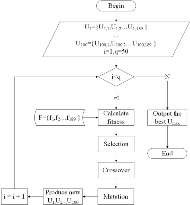
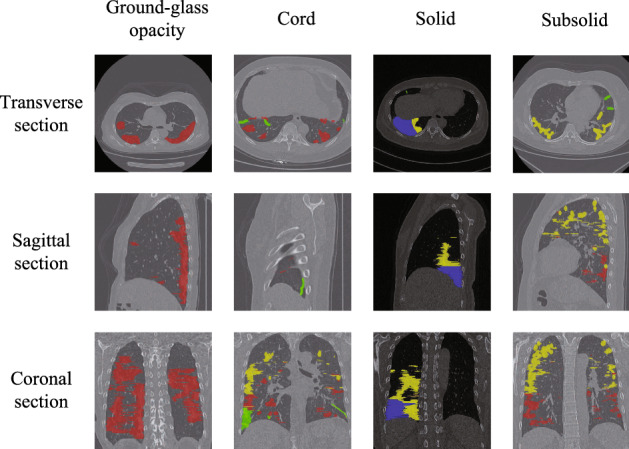
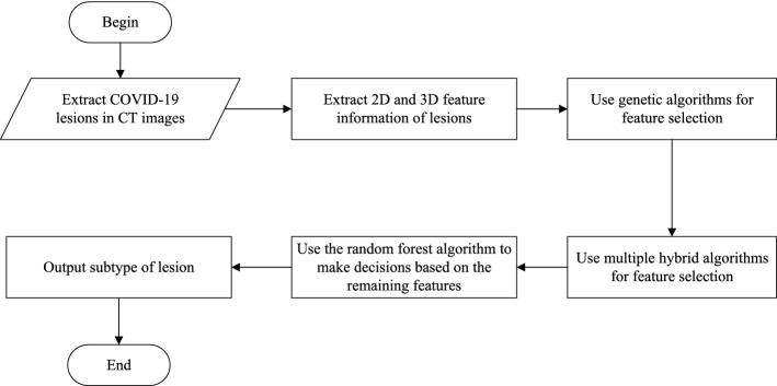
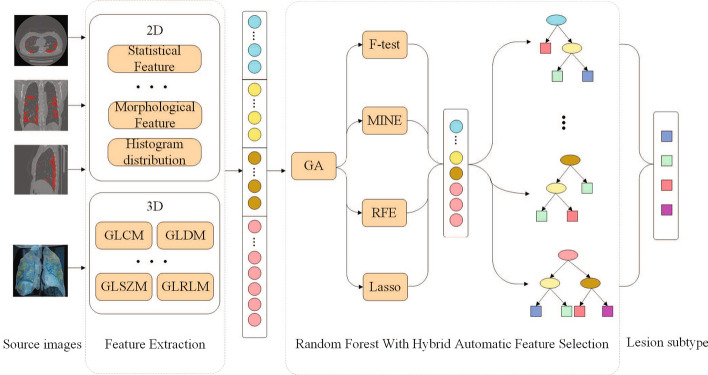
The most four typical lesion subtypes of COVID-19 are discussed in this paper, which are GGO (ground-glass opacity), cord, solid and subsolid. A computer-aided diagnosis approach of lesion subtype is proposed in this paper. The radiomics data of lesions are segmented from COVID-19 patients CT images with diagnosis and lesions annotations by radiologists. Then the three-dimensional texture descriptors are applied on the volume data of lesions as well as shape and first-order features. The massive feature data are selected by HAFS (hybrid adaptive feature selection) algorithm and a classification model is trained at the same time. The classifier is used to predict lesion subtypes as side decision information for radiologists.
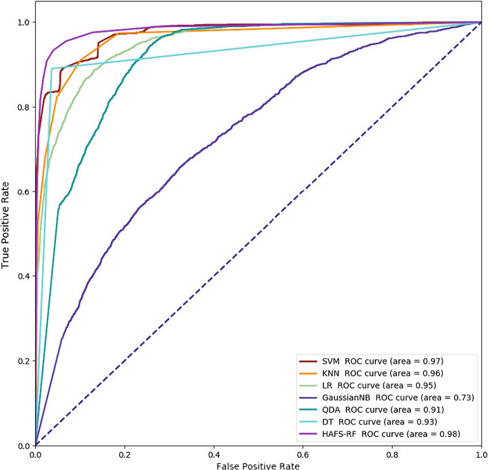
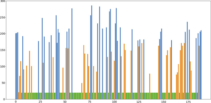
There are 3734 lesions extracted from the dataset with 319 patients collection and then 189 radiomics features are obtained finally. The random forest classifier is trained with data augmentation that the number of different subtypes of lesions is imbalanced in initial dataset. The experimental results show that the accuracy of the four subtypes of lesions is (93.06%, 96.84%, 99.58%, and 94.30%), the recall is (95.52%, 91.58%, 95.80% and 80.75%) and the f-score is (93.84%, 92.37%, 95.47%, and 84.42%).
The three-dimensional radiomics features used in this paper can better express the high-level information of COVID-19 lesions in CT slices. HAFS method aggregates the results of multiple feature selection algorithms intersects with traditional methods to filter out redundant features more accurately. After selection, the subtype of COVID-19 lesion can be judged by inputting the features into the RF (random forest) model, which can help clinicians more accurately identify the subtypes of COVID-19 lesions and provide help for further research.
COVID-19 疾病给全球医疗体系带来了前所未有的压力。CT(计算机断层扫描)检查作为一种辅助确诊方法,可以帮助临床医生在通过 PCR 检测进行筛查后,快速检测 COVID-19 的病变位置。此外,病变亚型分类在后续治疗决策中起着关键作用。准确识别病变亚型有助于医生及时发现病变变化,并更好地评估 COVID-19 的严重程度。
本文讨论了 COVID-19 最常见的四种典型病变亚型,即磨玻璃影(GGO)、条索影、实变和亚实性。本文提出了一种基于计算机辅助诊断的病变亚型分类方法。通过放射科医生对诊断和病变标注的 CT 图像,从 COVID-19 患者的 CT 图像中分割病变的放射组学数据。然后将三维纹理描述符应用于病变的体积数据以及形状和一阶特征。使用 HAFS(混合自适应特征选择)算法选择大量特征数据,并同时训练分类模型。该分类器用于预测病变亚型,作为放射科医生的辅助决策信息。
从 319 名患者的数据集中共提取了 3734 个病变,最终获得了 189 个放射组学特征。通过数据扩充,对初始数据集中小病变亚型数量不平衡的情况进行随机森林分类器训练。实验结果表明,四种病变亚型的准确率分别为(93.06%、96.84%、99.58%和 94.30%),召回率分别为(95.52%、91.58%、95.80%和 80.75%),F1 分数分别为(93.84%、92.37%、95.47%和 84.42%)。
本文使用的三维放射组学特征可以更好地表达 CT 切片中 COVID-19 病变的高级信息。HAFS 方法将多个特征选择算法的结果进行聚合,与传统方法交叉,更准确地筛选出冗余特征。选择后,通过将特征输入 RF(随机森林)模型,可以判断 COVID-19 病变的亚型,有助于临床医生更准确地识别 COVID-19 病变的亚型,为进一步研究提供帮助。