• 文献检索
  • 文档翻译
  • 深度研究
  • 学术资讯
  • Suppr Zotero 插件Zotero 插件
  • 邀请有礼
  • 套餐&价格
  • 历史记录
应用&插件
Suppr Zotero 插件Zotero 插件浏览器插件Mac 客户端Windows 客户端微信小程序
定价
高级版会员购买积分包购买API积分包
服务
文献检索文档翻译深度研究API 文档MCP 服务
关于我们
关于 Suppr公司介绍联系我们用户协议隐私条款
关注我们

Suppr 超能文献

核心技术专利:CN118964589B侵权必究
粤ICP备2023148730 号-1Suppr @ 2026

文献检索

告别复杂PubMed语法,用中文像聊天一样搜索,搜遍4000万医学文献。AI智能推荐,让科研检索更轻松。

立即免费搜索

文件翻译

保留排版,准确专业,支持PDF/Word/PPT等文件格式,支持 12+语言互译。

免费翻译文档

深度研究

AI帮你快速写综述,25分钟生成高质量综述,智能提取关键信息,辅助科研写作。

立即免费体验

基于影像组学数据的动态对比增强乳腺磁共振成像表型对乳腺癌分子亚型的识别

Molecular Subtypes Recognition of Breast Cancer in Dynamic Contrast-Enhanced Breast Magnetic Resonance Imaging Phenotypes from Radiomics Data.

机构信息

Key Laboratory of Intelligent Computing in Medical Image (MIIC), Northeastern University, Ministry of Education, Shenyang, China.

Biomedical and Information Engineering School, Northeastern University, Shenyang, China.

出版信息

Comput Math Methods Med. 2019 Oct 30;2019:6978650. doi: 10.1155/2019/6978650. eCollection 2019.

DOI:10.1155/2019/6978650
PMID:31827586
原文链接:https://pmc.ncbi.nlm.nih.gov/articles/PMC6885255/
Abstract

BACKGROUND AND OBJECTIVE

Breast cancer is a major cause of mortality among women if not treated in early stages. Recognizing molecular markers from DCE-MRI directly to distinguish the four molecular subtypes without invasive biopsy is helpful for guiding treatment plans for breast cancer, which provides a fast way to consequential treatment plan decision in early time and best opportunity for patients.

METHODS

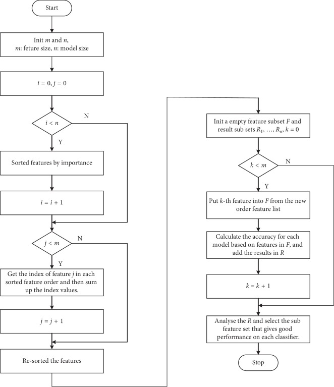
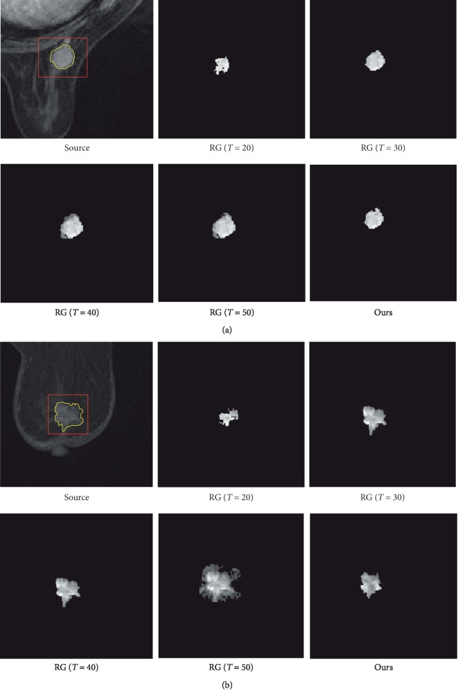
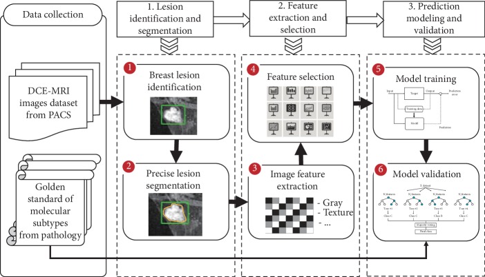
This study presents an approach of molecular subtypes recognition from breast cancer image phenotypes by radiomics. An improved region growth algorithm with dynamic threshold without user interaction is proposed for cancer lesion segmentation, which gives the precise border of lesion other than area with background. The lesions are extracted automatically based on radiologists' annotation which guarantees the lesion is segmented correctly. Various features are extracted on lesions data including texture, morphology, dynamic kinetics, and statistics features carried out on a large patient cohort, which are used to validate the relationship between image phenotypes and the molecular subtypes. A new algorithm of multimodel-based recursive feature elimination is applied on the radiomics data generated by the feature extraction process. This method obtains the feature subset with stable performance for different classification models, and the gradient boosting decision tree model gets the best results of both classification performance and imbalance performance on molecular subtypes.

RESULT

From the experimental results, 69 optimal features from 143 original features are found by the multimodel-based recursive feature elimination algorithms and the gradient boosting decision tree classifier obtains a good performance with accuracy 0.87, precise 0.88, recall 0.87, and F1-score 0.87. The dataset with 637 patients in this paper has serious imbalance problem on different molecular subtypes, and the the robust features that are generated by multimodel-based recursive feature eliminiation algorithm make the gradient boosting decision tree classifier have good behaviors. The recognition precision for the four molecular subtypes of luminal A, luminal B, HER-2, and basal-like are 0.91, 0.89, 0.83, and 0.87, respectively.

CONCLUSIONS

The improved lesion segmentation method gives more precise lesion edge, which not only saves the time of automatic extraction of lesion region of interest without threshold setting for each case, but also prevents the segmentation error by manual and prejudice from different radiologists. The feature selection algorithm of multimodel-based recursive feature elimination has the ability to find robust and optimal features that distinguish the four molecular subtypes from image phenotypes. The gradient boosting decision tree classifier rather plays a main role in recognition than other models used in this paper.

https://cdn.ncbi.nlm.nih.gov/pmc/blobs/2788/6885255/69224ba28397/CMMM2019-6978650.003.jpg
https://cdn.ncbi.nlm.nih.gov/pmc/blobs/2788/6885255/2e0b8ca7282e/CMMM2019-6978650.001.jpg
https://cdn.ncbi.nlm.nih.gov/pmc/blobs/2788/6885255/69033b40dbf4/CMMM2019-6978650.002.jpg
https://cdn.ncbi.nlm.nih.gov/pmc/blobs/2788/6885255/69224ba28397/CMMM2019-6978650.003.jpg
https://cdn.ncbi.nlm.nih.gov/pmc/blobs/2788/6885255/2e0b8ca7282e/CMMM2019-6978650.001.jpg
https://cdn.ncbi.nlm.nih.gov/pmc/blobs/2788/6885255/69033b40dbf4/CMMM2019-6978650.002.jpg
https://cdn.ncbi.nlm.nih.gov/pmc/blobs/2788/6885255/69224ba28397/CMMM2019-6978650.003.jpg
摘要

背景与目的

如果不在早期阶段进行治疗,乳腺癌是导致女性死亡的主要原因之一。直接从 DCE-MRI 中识别分子标志物来区分四种分子亚型,而无需进行侵入性活检,有助于指导乳腺癌的治疗计划,为患者提供早期及时的治疗计划决策和最佳机会。

方法

本研究通过放射组学从乳腺癌图像表型中提出一种识别分子亚型的方法。提出了一种改进的区域生长算法,该算法具有无需用户交互的动态阈值,可用于癌症病变分割,该算法可以提供病变的精确边界,而不仅仅是背景区域的面积。病变是基于放射科医生的注释自动提取的,这保证了病变的正确分割。在大型患者队列中对病变数据进行了各种特征提取,包括纹理、形态、动态动力学和统计特征,用于验证图像表型与分子亚型之间的关系。应用一种新的多模型递归特征消除算法对特征提取过程生成的放射组学数据进行处理。该方法获得了具有不同分类模型稳定性能的特征子集,梯度提升决策树模型在分子亚型的分类性能和不平衡性能方面均取得了最佳结果。

结果

从实验结果可以看出,通过多模型递归特征消除算法找到了 69 个最优特征,梯度提升决策树分类器的性能良好,准确率为 0.87,精确率为 0.88,召回率为 0.87,F1 得分为 0.87。本文数据集有 637 名患者,不同分子亚型之间存在严重的不平衡问题,多模型递归特征消除算法生成的稳健特征使梯度提升决策树分类器具有良好的性能。四种分子亚型(管腔 A、管腔 B、HER-2 和基底样)的识别精度分别为 0.91、0.89、0.83 和 0.87。

结论

改进的病变分割方法提供了更精确的病变边缘,不仅节省了为每个病例设置阈值自动提取病变感兴趣区的时间,而且还防止了不同放射科医生的手动和偏见造成的分割错误。多模型递归特征消除的特征选择算法具有从图像表型中找到区分四种分子亚型的稳健和最优特征的能力。梯度提升决策树分类器在识别中起着比本文中使用的其他模型更重要的作用。

相似文献

1
Molecular Subtypes Recognition of Breast Cancer in Dynamic Contrast-Enhanced Breast Magnetic Resonance Imaging Phenotypes from Radiomics Data.基于影像组学数据的动态对比增强乳腺磁共振成像表型对乳腺癌分子亚型的识别
Comput Math Methods Med. 2019 Oct 30;2019:6978650. doi: 10.1155/2019/6978650. eCollection 2019.
2
Textural kinetics: a novel dynamic contrast-enhanced (DCE)-MRI feature for breast lesion classification.纹理动力学:一种新的动态对比增强(DCE)MRI 特征,用于乳腺病变分类。
J Digit Imaging. 2011 Jun;24(3):446-63. doi: 10.1007/s10278-010-9298-1.
3
Multilevel analysis of spatiotemporal association features for differentiation of tumor enhancement patterns in breast DCE-MRI.基于多时相分析的乳腺 DCE-MRI 肿瘤增强模式鉴别特征研究。
Med Phys. 2010 Aug;37(8):3940-56. doi: 10.1118/1.3446799.
4
Malignant lesion segmentation in contrast-enhanced breast MR images based on the marker-controlled watershed.基于标记控制分水岭的对比增强乳腺磁共振图像中的恶性病变分割。
Med Phys. 2009 Oct;36(10):4359-69. doi: 10.1118/1.3213514.
5
Automatic identification and classification of characteristic kinetic curves of breast lesions on DCE-MRI.动态对比增强磁共振成像(DCE-MRI)中乳腺病变特征性动力学曲线的自动识别与分类
Med Phys. 2006 Aug;33(8):2878-87. doi: 10.1118/1.2210568.
6
A quantitative heterogeneity analysis approach to molecular subtype recognition of breast cancer in dynamic contrast-enhanced magnetic imaging images from radiomics data.一种基于放射组学数据的动态对比增强磁共振成像图像中乳腺癌分子亚型识别的定量异质性分析方法。
Quant Imaging Med Surg. 2023 Jul 1;13(7):4429-4446. doi: 10.21037/qims-22-1230. Epub 2023 May 26.
7
Radiomic analysis reveals DCE-MRI features for prediction of molecular subtypes of breast cancer.放射组学分析揭示了用于预测乳腺癌分子亚型的动态对比增强磁共振成像(DCE-MRI)特征。
PLoS One. 2017 Feb 6;12(2):e0171683. doi: 10.1371/journal.pone.0171683. eCollection 2017.
8
Spectral embedding based active contour (SEAC) for lesion segmentation on breast dynamic contrast enhanced magnetic resonance imaging.基于谱嵌入的主动轮廓(SEAC)用于乳腺动态对比增强磁共振成像的病变分割。
Med Phys. 2013 Mar;40(3):032305. doi: 10.1118/1.4790466.
9
Radiomic Signatures Derived from Diffusion-Weighted Imaging for the Assessment of Breast Cancer Receptor Status and Molecular Subtypes.基于扩散加权成像的影像组学特征评估乳腺癌受体状态和分子亚型。
Mol Imaging Biol. 2020 Apr;22(2):453-461. doi: 10.1007/s11307-019-01383-w.
10
A new automated method for the segmentation and characterization of breast masses on ultrasound images.一种用于超声图像上乳腺肿块分割与特征描述的新型自动化方法。
Med Phys. 2009 May;36(5):1553-65. doi: 10.1118/1.3110069.

引用本文的文献

1
Radiomics Integration of Mammography and DCE-MRI for Predicting Molecular Subtypes in Breast Cancer Patients.乳腺钼靶和动态对比增强磁共振成像的影像组学整合用于预测乳腺癌患者的分子亚型
Breast Cancer (Dove Med Press). 2025 Feb 18;17:187-200. doi: 10.2147/BCTT.S488200. eCollection 2025.
2
The effect of data resampling methods in radiomics.数据重采样方法在放射组学中的影响。
Sci Rep. 2024 Feb 3;14(1):2858. doi: 10.1038/s41598-024-53491-5.
3
Breast Cancer Surrogate Subtype Classification Using Pretreatment Multi-Phase Dynamic Contrast-Enhanced Magnetic Resonance Imaging Radiomics: A Retrospective Single-Center Study.

本文引用的文献

1
Deep learning for identifying radiogenomic associations in breast cancer.深度学习在乳腺癌放射基因组关联识别中的应用。
Comput Biol Med. 2019 Jun;109:85-90. doi: 10.1016/j.compbiomed.2019.04.018. Epub 2019 Apr 25.
2
Predicting Breast Cancer Molecular Subtype with MRI Dataset Utilizing Convolutional Neural Network Algorithm.利用卷积神经网络算法对 MRI 数据集进行乳腺癌分子亚型预测。
J Digit Imaging. 2019 Apr;32(2):276-282. doi: 10.1007/s10278-019-00179-2.
3
Breast DCE-MRI: lesion classification using dynamic and morphological features by means of a multiple classifier system.
利用治疗前多期动态对比增强磁共振成像放射组学进行乳腺癌替代亚型分类:一项回顾性单中心研究
J Pers Med. 2023 Jul 18;13(7):1150. doi: 10.3390/jpm13071150.
4
A quantitative heterogeneity analysis approach to molecular subtype recognition of breast cancer in dynamic contrast-enhanced magnetic imaging images from radiomics data.一种基于放射组学数据的动态对比增强磁共振成像图像中乳腺癌分子亚型识别的定量异质性分析方法。
Quant Imaging Med Surg. 2023 Jul 1;13(7):4429-4446. doi: 10.21037/qims-22-1230. Epub 2023 May 26.
5
Prediction of distant metastasis in esophageal cancer using a radiomics-clinical model.基于放射组学-临床模型预测食管癌远处转移。
Eur J Med Res. 2022 Dec 3;27(1):272. doi: 10.1186/s40001-022-00877-8.
6
Invasive ductal breast cancer molecular subtype prediction by MRI radiomic and clinical features based on machine learning.基于机器学习,通过MRI影像组学和临床特征预测浸润性导管乳腺癌分子亚型
Front Oncol. 2022 Sep 12;12:964605. doi: 10.3389/fonc.2022.964605. eCollection 2022.
7
Radiomic Signatures Derived from Hybrid Contrast-Enhanced Ultrasound Images (CEUS) for the Assessment of Histological Characteristics of Breast Cancer: A Pilot Study.基于混合对比增强超声图像(CEUS)的放射组学特征用于评估乳腺癌组织学特征的初步研究
Cancers (Basel). 2022 Aug 12;14(16):3905. doi: 10.3390/cancers14163905.
8
Influence of Percutaneous Drainage Surgery and the Interval to Perform Laparoscopic Cholecystectomy on Acute Cholecystitis through Genetic Algorithm-Based Contrast-Enhanced Ultrasound Imaging.基于遗传算法的对比增强超声成像对经皮引流术及行腹腔镜胆囊切除术时间间隔对急性胆囊炎的影响。
Comput Intell Neurosci. 2022 Jul 30;2022:3602811. doi: 10.1155/2022/3602811. eCollection 2022.
9
Breast Cancer Classification on Multiparametric MRI - Increased Performance of Boosting Ensemble Methods.多参数 MRI 乳腺癌分类-提升集成方法的性能。
Technol Cancer Res Treat. 2022 Jan-Dec;21:15330338221087828. doi: 10.1177/15330338221087828.
10
Co-clinical FDG-PET radiomic signature in predicting response to neoadjuvant chemotherapy in triple-negative breast cancer.三阴性乳腺癌新辅助化疗疗效的 co-clinical FDG-PET 影像组学特征。
Eur J Nucl Med Mol Imaging. 2022 Jan;49(2):550-562. doi: 10.1007/s00259-021-05489-8. Epub 2021 Jul 30.
乳腺动态对比增强磁共振成像:通过多分类器系统利用动态和形态学特征进行病变分类
Eur Radiol Exp. 2017;1(1):10. doi: 10.1186/s41747-017-0007-4. Epub 2017 Jun 29.
4
Breast MRI radiomics: comparison of computer- and human-extracted imaging phenotypes.乳腺MRI影像组学:计算机提取与人工提取影像表型的比较
Eur Radiol Exp. 2017;1(1):22. doi: 10.1186/s41747-017-0025-2. Epub 2017 Nov 21.
5
Spatiotemporal features of DCE-MRI for breast cancer diagnosis.用于乳腺癌诊断的 DCE-MRI 的时空特征。
Comput Methods Programs Biomed. 2018 Mar;155:153-164. doi: 10.1016/j.cmpb.2017.12.015. Epub 2017 Dec 12.
6
MRI texture analysis in differentiating luminal A and luminal B breast cancer molecular subtypes - a feasibility study.磁共振成像纹理分析在鉴别管腔A型和管腔B型乳腺癌分子亚型中的应用——一项可行性研究
BMC Med Imaging. 2017 Dec 29;17(1):69. doi: 10.1186/s12880-017-0239-z.
7
Radiomic analysis of DCE-MRI for prediction of response to neoadjuvant chemotherapy in breast cancer patients.基于 DCE-MRI 的影像组学分析预测乳腺癌患者新辅助化疗的疗效。
Eur J Radiol. 2017 Sep;94:140-147. doi: 10.1016/j.ejrad.2017.06.019. Epub 2017 Jun 28.
8
DCE-MRI Texture Features for Early Prediction of Breast Cancer Therapy Response.用于早期预测乳腺癌治疗反应的动态对比增强磁共振成像纹理特征
Tomography. 2017 Mar;3(1):23-32. doi: 10.18383/j.tom.2016.00241.
9
Maximum intensity breast diffusion MRI for BI-RADS 4 lesions detected on X-ray mammography.针对乳腺X线摄影检测出的BI-RADS 4类病变的最大强度乳腺扩散磁共振成像
Clin Radiol. 2017 Oct;72(10):900.e1-900.e8. doi: 10.1016/j.crad.2017.05.017. Epub 2017 Jul 4.
10
Intratumoral and peritumoral radiomics for the pretreatment prediction of pathological complete response to neoadjuvant chemotherapy based on breast DCE-MRI.基于乳腺动态对比增强磁共振成像的瘤内和瘤周影像组学对新辅助化疗病理完全缓解的治疗前预测
Breast Cancer Res. 2017 May 18;19(1):57. doi: 10.1186/s13058-017-0846-1.