• 文献检索
  • 文档翻译
  • 深度研究
  • 学术资讯
  • Suppr Zotero 插件Zotero 插件
  • 邀请有礼
  • 套餐&价格
  • 历史记录
应用&插件
Suppr Zotero 插件Zotero 插件浏览器插件Mac 客户端Windows 客户端微信小程序
定价
高级版会员购买积分包购买API积分包
服务
文献检索文档翻译深度研究API 文档MCP 服务
关于我们
关于 Suppr公司介绍联系我们用户协议隐私条款
关注我们

Suppr 超能文献

核心技术专利:CN118964589B侵权必究
粤ICP备2023148730 号-1Suppr @ 2026

文献检索

告别复杂PubMed语法,用中文像聊天一样搜索,搜遍4000万医学文献。AI智能推荐,让科研检索更轻松。

立即免费搜索

文件翻译

保留排版,准确专业,支持PDF/Word/PPT等文件格式,支持 12+语言互译。

免费翻译文档

深度研究

AI帮你快速写综述,25分钟生成高质量综述,智能提取关键信息,辅助科研写作。

立即免费体验

病例报告:新型冠状病毒肺炎后的大疱性肺疾病

Case Report: Bullous Lung Disease Following COVID-19.

作者信息

Pednekar Prachi, Amoah Kwesi, Homer Robert, Ryu Changwan, Lutchmansingh Denyse D

机构信息

Department of Internal Medicine, Bridgeport Hospital, Bridgeport, CT, United States.

Section of Pulmonary, Critical Care, and Sleep Medicine, Bridgeport Hospital, Bridgeport, CT, United States.

出版信息

Front Med (Lausanne). 2021 Nov 17;8:770778. doi: 10.3389/fmed.2021.770778. eCollection 2021.

DOI:10.3389/fmed.2021.770778
PMID:34869488
原文链接:https://pmc.ncbi.nlm.nih.gov/articles/PMC8635639/
Abstract

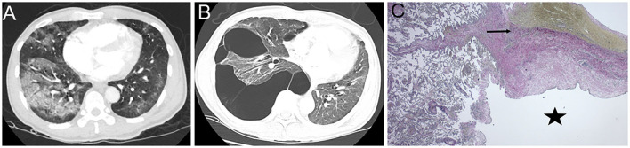
More than 87% of patients report the persistence of at least one symptom after recovery from the Coronavirus disease 2019 (COVID-19). Dyspnea is one of the most frequently reported symptoms following severe acute respiratory syndrome coronavirus-2 (SARS CoV-2) infection with persistent chest radiological abnormalities up to 3 months after symptom onset. These radiological abnormalities are variable and most commonly include ground-glass opacities, reticulations, mosaic attenuation, parenchymal bands, interlobular septal thickening, bronchiectasis, and fibrotic-like changes. However, in this case report, we describe findings of bullous lung disease as a complication of SARS CoV-2 infection. As the pandemic continues, there is a need to understand the multiple respiratory manifestations of post-acute sequelae of COVID-19. We, therefore, present this case to add to the current body of literature describing pulmonary disease as a consequence of SARS CoV-2 infection.

摘要

超过87%的患者报告称,在从2019冠状病毒病(COVID-19)康复后至少有一种症状持续存在。呼吸困难是严重急性呼吸综合征冠状病毒2(SARS-CoV-2)感染后最常报告的症状之一,症状出现后长达3个月胸部影像学异常持续存在。这些影像学异常多种多样,最常见的包括磨玻璃影、网状影、马赛克样衰减、实质带、小叶间隔增厚、支气管扩张和纤维化样改变。然而,在本病例报告中,我们描述了大疱性肺病作为SARS-CoV-2感染并发症的表现。随着疫情的持续,有必要了解COVID-19急性后遗症的多种呼吸道表现。因此,我们呈现此病例,以补充目前描述SARS-CoV-2感染所致肺部疾病的文献。

https://cdn.ncbi.nlm.nih.gov/pmc/blobs/eb03/8635639/c226260fac3a/fmed-08-770778-g0001.jpg

相似文献

1
Case Report: Bullous Lung Disease Following COVID-19.病例报告:新型冠状病毒肺炎后的大疱性肺疾病
Front Med (Lausanne). 2021 Nov 17;8:770778. doi: 10.3389/fmed.2021.770778. eCollection 2021.
2
Chest CT Findings at Six Months Following COVID-19 ARDS - Correlation With the mMRC Dyspnea Scale and Pulmonary Function Tests.COVID-19 后 ARDS 患者 6 个月时的胸部 CT 表现与 mMRC 呼吸困难量表和肺功能测试的相关性。
Br J Biomed Sci. 2024 Jul 10;81:12871. doi: 10.3389/bjbs.2024.12871. eCollection 2024.
3
Chest CT Lung Abnormalities 1 Year after COVID-19: A Systematic Review and Meta-Analysis.COVID-19 后 1 年的胸部 CT 肺部异常:系统评价和荟萃分析。
Radiology. 2023 Jul;308(1):e230535. doi: 10.1148/radiol.230535.
4
Short- and Long-Term Chest-CT Findings after Recovery from COVID-19: A Systematic Review and Meta-Analysis.新冠康复后的短期和长期胸部CT表现:一项系统评价和荟萃分析
Diagnostics (Basel). 2024 Mar 14;14(6):621. doi: 10.3390/diagnostics14060621.
5
Imaging and clinical features of patients with 2019 novel coronavirus SARS-CoV-2.新型冠状病毒 2019 年 SARS-CoV-2 患者的影像学和临床特征。
Eur J Nucl Med Mol Imaging. 2020 May;47(5):1275-1280. doi: 10.1007/s00259-020-04735-9. Epub 2020 Feb 28.
6
[Follow-up of patients after COVID-19 pneumonia. Pulmonary sequelae].新型冠状病毒肺炎后患者的随访。肺部后遗症
Rev Alerg Mex. 2020 Oct-Dec;67(4):350-369. doi: 10.29262/ram.v67i4.847.
7
[Morphological and functional sequelae after COVID-19 pneumonia].[新型冠状病毒肺炎后的形态学和功能后遗症]
Radiologe. 2021 Oct;61(10):888-895. doi: 10.1007/s00117-021-00905-4. Epub 2021 Sep 16.
8
Chest CT in covid-19 pneumonia's follow-up: A 30 patients case series.新型冠状病毒肺炎随访中的胸部CT:30例病例系列报道
Ann Med Surg (Lond). 2022 Dec;84:104835. doi: 10.1016/j.amsu.2022.104835. Epub 2022 Nov 7.
9
COVID-19 pneumonia: CT findings of 122 patients and differentiation from influenza pneumonia.新型冠状病毒肺炎:122例患者的CT表现及与流感肺炎的鉴别诊断
Eur Radiol. 2020 Oct;30(10):5463-5469. doi: 10.1007/s00330-020-06928-0. Epub 2020 May 12.
10
[Pulmonary manifestations in long COVID].[新冠长期症状中的肺部表现]
Inn Med (Heidelb). 2022 Aug;63(8):819-829. doi: 10.1007/s00108-022-01371-3. Epub 2022 Jul 7.

引用本文的文献

1
A Case of Interstitial Pneumonia Leading to Respiratory Failure Several Months After COVID-19 Infection.1例新型冠状病毒肺炎感染数月后导致呼吸衰竭的间质性肺炎病例。
Cureus. 2025 Jan 8;17(1):e77153. doi: 10.7759/cureus.77153. eCollection 2025 Jan.
2
Bullous lung disease in pregnancy: a case report.妊娠期间的大疱性肺病:一例报告。
Obstet Med. 2024 Jul 26;18(3):190-192. doi: 10.1177/1753495X241265528. eCollection 2025 Sep.
3
Paraseptal Lucencies Mimicking Emphysema in a Non-smoker With Acute Lung Injury in COVID-19.新冠病毒肺炎急性肺损伤非吸烟者中类似肺气肿的肺小叶间隔透亮区

本文引用的文献

1
A case of bulla formation after treatment for COVID-19 pneumonia.1例新型冠状病毒肺炎治疗后出现大疱形成的病例。
Radiol Case Rep. 2021 May;16(5):1162-1164. doi: 10.1016/j.radcr.2021.03.003. Epub 2021 Mar 10.
2
The pulmonary pathology of COVID-19.COVID-19 的肺部病理学。
Virchows Arch. 2021 Jan;478(1):137-150. doi: 10.1007/s00428-021-03053-1. Epub 2021 Feb 19.
3
Pulmonary emphysema, bullae, and pneumothorax in COVID-19 pneumonia.新型冠状病毒肺炎中的肺气肿、肺大疱和气胸。
Cureus. 2024 Oct 7;16(10):e71010. doi: 10.7759/cureus.71010. eCollection 2024 Oct.
4
Bullous Lung Disease due to Pulmonary Tuberculosis: A Rare Case Complicated With Tension Pneumothorax and Bronchopleural Fistula.肺结核所致大疱性肺病:1例合并张力性气胸和支气管胸膜瘘的罕见病例
Ther Adv Pulm Crit Care Med. 2024 May 10;19:29768675241249652. doi: 10.1177/29768675241249652. eCollection 2024 Jan-Jun.
5
Critical Damage of Lung Parenchyma Complicated with Massive Pneumothorax in COVID-19 Pneumonia.新型冠状病毒肺炎肺实质严重损伤合并大量气胸
Clin Med Insights Case Rep. 2023 May 20;16:11795476231175644. doi: 10.1177/11795476231175644. eCollection 2023.
6
Development of bullous lung disease with pneumothorax following SARS-CoV-2 infection.新型冠状病毒感染后伴气胸的大疱性肺疾病的发展
Respirol Case Rep. 2022 Aug 2;10(9):e01013. doi: 10.1002/rcr2.1013. eCollection 2022 Sep.
Radiol Case Rep. 2021 May;16(5):995-998. doi: 10.1016/j.radcr.2021.01.055. Epub 2021 Jan 31.
4
Delayed recurrent spontaneous pneumothorax post-recovery from COVID-19 infection.新冠病毒感染康复后延迟复发的自发性气胸
Indian J Thorac Cardiovasc Surg. 2021 Sep;37(5):551-553. doi: 10.1007/s12055-021-01145-w. Epub 2021 Feb 2.
5
Six-month Follow-up Chest CT Findings after Severe COVID-19 Pneumonia.重症 COVID-19 肺炎患者 6 个月随访的胸部 CT 结果。
Radiology. 2021 Apr;299(1):E177-E186. doi: 10.1148/radiol.2021203153. Epub 2021 Jan 26.
6
Hemoptysis and a Newly Formed Lung Bulla in a Case of Convalescent COVID-19 Pneumonia.一名新冠康复期肺炎患者出现咯血及新形成的肺大疱
Intern Med. 2021 Mar 1;60(5):803-805. doi: 10.2169/internalmedicine.5684-20. Epub 2021 Jan 15.
7
Persistent Post-COVID-19 Interstitial Lung Disease. An Observational Study of Corticosteroid Treatment.持续性 COVID-19 间质性肺病。皮质类固醇治疗的观察性研究。
Ann Am Thorac Soc. 2021 May;18(5):799-806. doi: 10.1513/AnnalsATS.202008-1002OC.
8
Pulmonary function and radiological features 4 months after COVID-19: first results from the national prospective observational Swiss COVID-19 lung study.新冠病毒病感染4个月后的肺功能和放射学特征:瑞士全国前瞻性观察性新冠病毒病肺部研究的初步结果
Eur Respir J. 2021 Apr 29;57(4). doi: 10.1183/13993003.03690-2020. Print 2021 Apr.
9
Imaging in the aftermath of COVID-19: what to expect.新冠疫情后的影像学表现:预期情况
Eur Radiol. 2021 Jun;31(6):4390-4392. doi: 10.1007/s00330-020-07465-6. Epub 2020 Dec 28.
10
Development of bullous lung disease in a patient with severe COVID-19 pneumonitis.一名重症新型冠状病毒肺炎患者发生大疱性肺疾病。
BMJ Case Rep. 2020 Oct 29;13(10):e237455. doi: 10.1136/bcr-2020-237455.