Murayama Daisuke, Kojima Daichi, Hino Ayako, Yamamoto Yayoi, Doiuchi Tsunehiro, Horikawa Ayumi, Kurihara Hiroaki
Department of Diagnostic and Interventional Radiology, Kanagawa Cancer Center, 2-3-2 Nakao Asahi-ku, Yokohama 241-8515, Kanagawa, Japan.
Radiol Case Rep. 2021 May;16(5):1162-1164. doi: 10.1016/j.radcr.2021.03.003. Epub 2021 Mar 10.
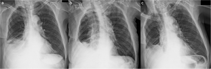
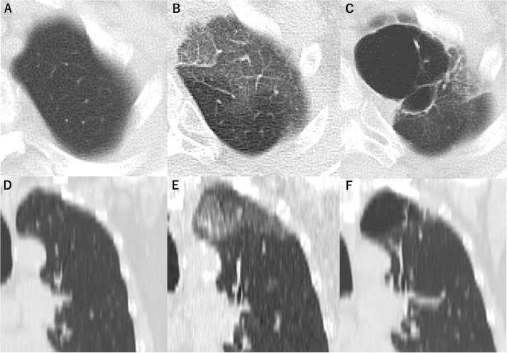
In December 2019, coronavirus disease 2019 (COVID-19), which is caused by severe acute respiratory syndrome coronavirus 2 (SARS-CoV-2), was reported in Wuhan, China. An 82-year-old woman presented to our hospital with high fever (39°C) and chest computed tomography revealed ground-glass opacities in the left lung apex. She was positive for SARS-CoV-2 based on a polymerase chain reaction test, and diagnosed with COVID-19 pneumonia. 6 months after treatment, chest CT showed a large bulla (47 mm × 29 mm) in the left lung apex, although pneumonia had partially resolved. Radiologic follow-up is needed after COVID-19 pneumonia, because patients may develop bullae after treatment.
2019年12月,中国武汉报告了由严重急性呼吸综合征冠状病毒2(SARS-CoV-2)引起的2019冠状病毒病(COVID-19)。一名82岁女性因高热(39°C)前来我院就诊,胸部计算机断层扫描显示左肺尖有磨玻璃影。基于聚合酶链反应检测,她的SARS-CoV-2呈阳性,并被诊断为COVID-19肺炎。治疗6个月后,尽管肺炎已部分消退,但胸部CT显示左肺尖有一个大疱(47 mm×29 mm)。COVID-19肺炎后需要进行影像学随访,因为患者在治疗后可能会出现肺大疱。