Ma Yanpeng, Zhang Yong, Qiu Chuan, He Chunhui, He Ting, Shi Shuang, Liu Zhongwei
Department of Cardiology, Shaanxi Provincial People's Hospital, Xi'an, China.
Center for Bioinformatics and Genomics, Department of Global Biostatistics and Data Science, School of Public Health and Tropical Medicine, Tulane University, New Orleans, LA, United States.
Front Cardiovasc Med. 2021 Nov 15;8:739212. doi: 10.3389/fcvm.2021.739212. eCollection 2021.
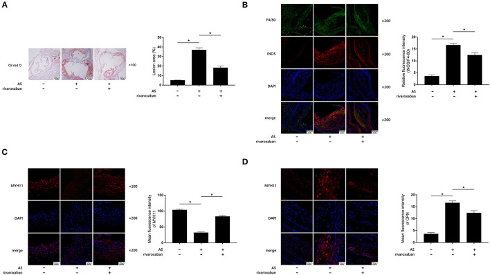
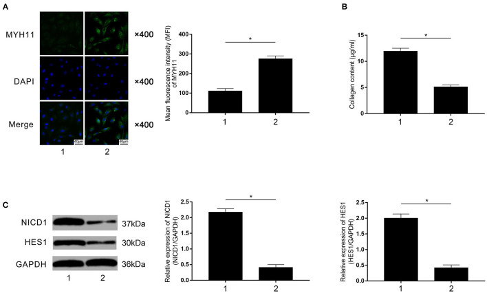
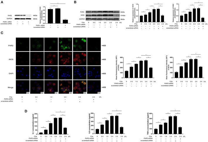
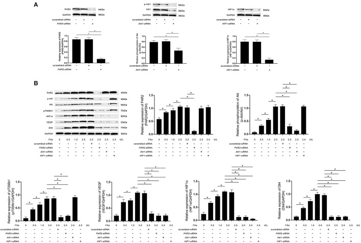
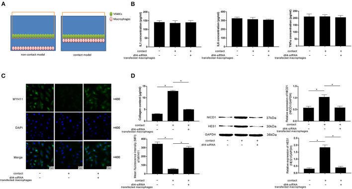
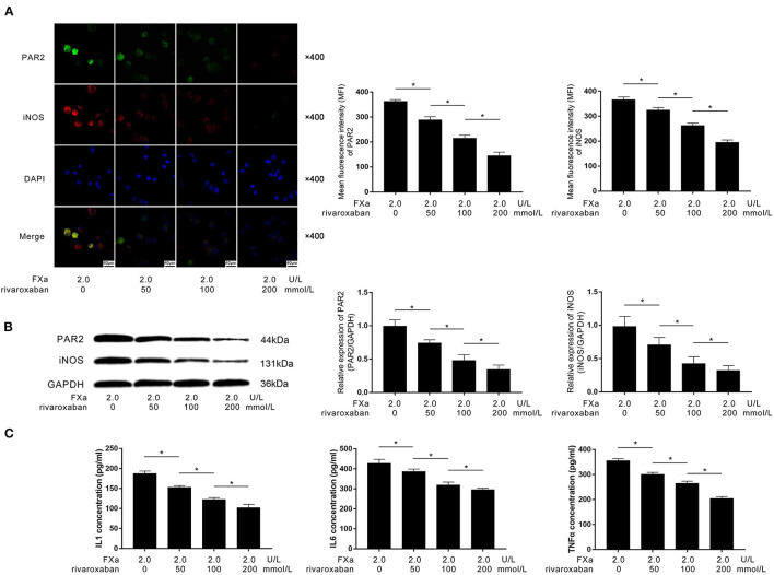
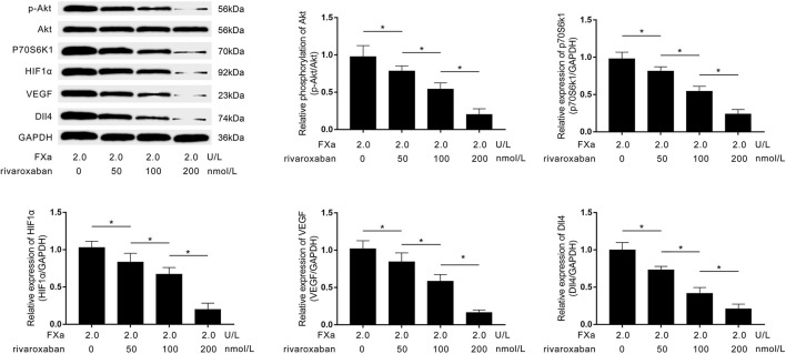
Factor Xa (FXa) is a mediator initiating and accelerating atherosclerosis (AS). Both macrophage and vascular smooth muscle cells (VSMCs) participate in AS progression. This study was aimed to investigate the mechanisms underlying the effects of the FXa inhibitor rivaroxaban on AS. Rivaroxaban was administered to AS mice. Primary macrophages were exposed to FXa, treated with rivaroxaban, and transfected with siRNA silencing protease-activated receptor 2 (PAR2), hypoxia-inducible factor 1α (HIF1α), delta-like receptor 4 (Dll4), and Akt. Interaction between macrophages and VSMCs was assessed by co-culturing systems. Atherosclerotic lesions were evaluated by oil red O stain. Fluorescent staining was used to determine the cell phenotypes. Secretions of inflammatory cytokines and collagen were assessed by ELISA and Sircol assays. Western blotting was used to evaluate the protein expression and phosphorylation levels. Rivaroxaban reduced lesion area, accumulation of M1 macrophages, and contractile-synthetic phenotypic conversion of VSMCs in atherosclerotic plaques. FXa exposure induced polarization of macrophages toward M1 and Dll4 high expression, which were inhibited by , and α silencing. Rivaroxaban treatment inhibited PAR2/Akt/HIF1α signaling activation and Dll4 expression in FXa-exposed macrophages. By cell-to-cell contact, M1 macrophages induced Notch signaling activation in VSMCs which committed contractile-synthetic conversion. Rivaroxaban treatment and Dll4 silencing incapacitated macrophage in inducing phenotypic conversion of VSMCs upon cell-to-cell contact. Rivaroxaban suppresses AS by inhibiting FXa-induced PAR2/Akt/HIF1α signaling activation-mediated macrophage M1 polarization and high Dll4 expression. These macrophages facilitated VSMCs to perform contractile-synthetic phenotypic conversion upon macrophage-VSMCs cell-to-cell contact.
凝血因子Xa(FXa)是引发和加速动脉粥样硬化(AS)的介质。巨噬细胞和血管平滑肌细胞(VSMC)均参与AS的进展。本研究旨在探讨FXa抑制剂利伐沙班对AS作用的潜在机制。将利伐沙班给予AS小鼠。原代巨噬细胞暴露于FXa,用利伐沙班处理,并转染沉默蛋白酶激活受体2(PAR2)、缺氧诱导因子1α(HIF1α)、Delta样受体4(Dll4)和Akt的小干扰RNA(siRNA)。通过共培养系统评估巨噬细胞与VSMC之间的相互作用。通过油红O染色评估动脉粥样硬化病变。使用荧光染色确定细胞表型。通过酶联免疫吸附测定(ELISA)和Sircol检测评估炎性细胞因子和胶原蛋白的分泌。使用蛋白质免疫印迹法评估蛋白质表达和磷酸化水平。利伐沙班减少了动脉粥样硬化斑块中的病变面积、M1巨噬细胞的积聚以及VSMC的收缩-合成表型转化。FXa暴露诱导巨噬细胞向M1极化和Dll4高表达,而PAR2、HIF1α和Akt沉默可抑制这种极化和高表达。利伐沙班处理抑制了FXa暴露的巨噬细胞中PAR2/Akt/HIF1α信号激活和Dll4表达。通过细胞间接触,M1巨噬细胞诱导VSMC中的Notch信号激活,从而导致收缩-合成转化。利伐沙班处理和Dll4沉默使巨噬细胞在细胞间接触时无法诱导VSMC的表型转化。利伐沙班通过抑制FXa诱导的PAR2/Akt/HIF1α信号激活介导的巨噬细胞M1极化和高Dll4表达来抑制AS。这些巨噬细胞在巨噬细胞与VSMC细胞间接触时促进VSMC进行收缩-合成表型转化。