Department of Orthopaedics, Jinling Hospital, School of Medicine, Nanjing University, Nanjing, China.
Department of Orthopaedics, The First Affiliated Hospital of Bengbu Medical College, Laboratory of Tissue and Transplant in Anhui Province, Bengbu Medical College, Bengbu, China.
Orthop Surg. 2022 Jan;14(1):119-128. doi: 10.1111/os.13183. Epub 2021 Dec 13.
To develop a new method to restore hip rotation center exactly and rapidly in total hip arthroplasty (THA) with the assistance of three dimensional (3D) printing technology and evaluate its clinical and radiological outcomes.
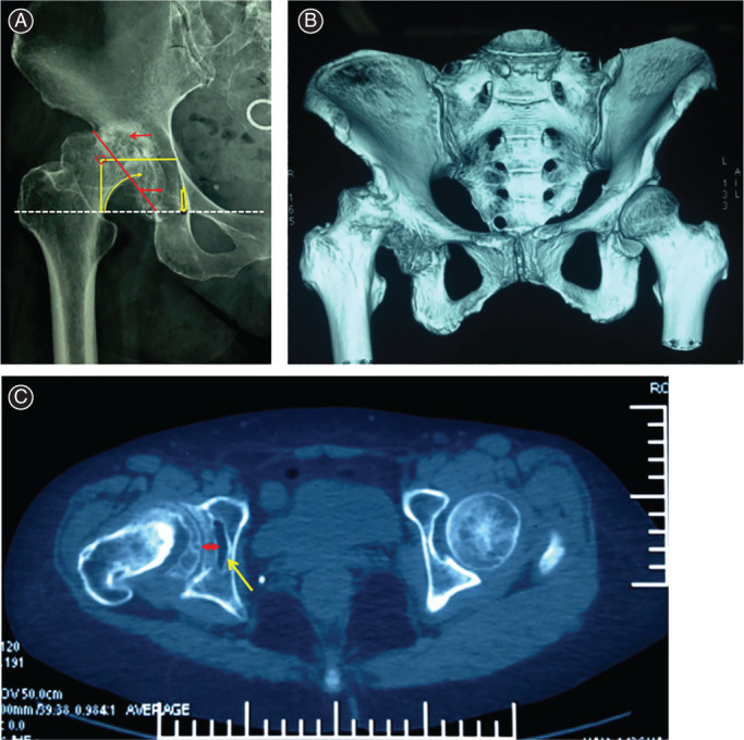
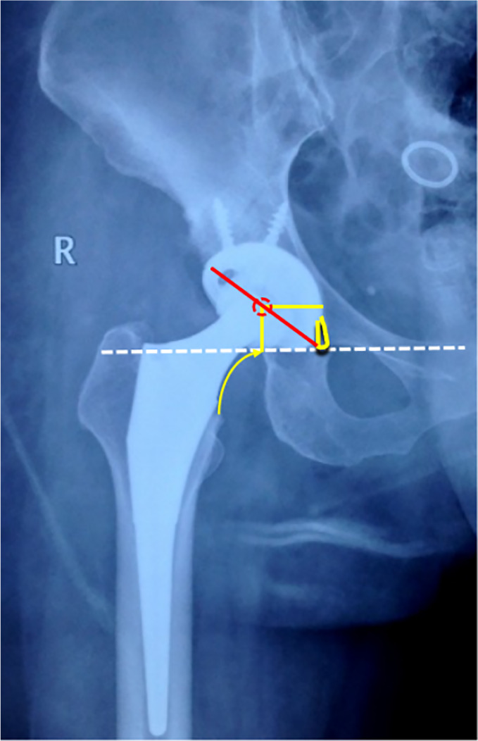
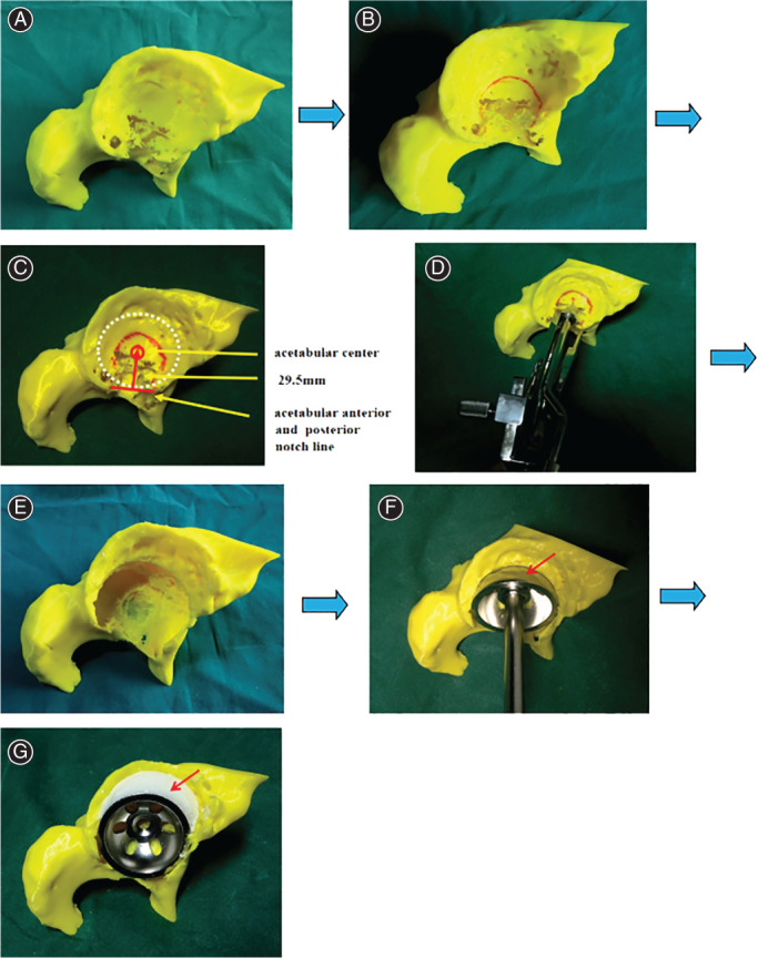
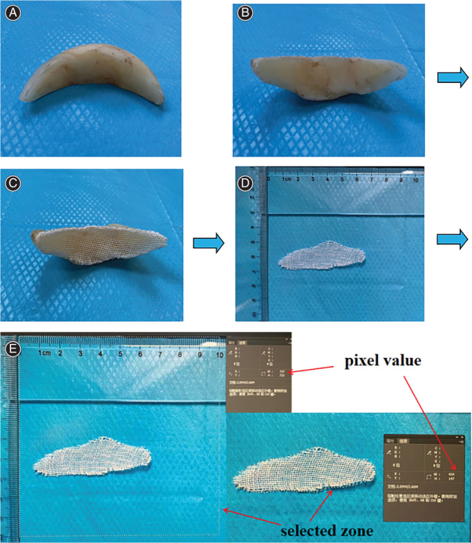
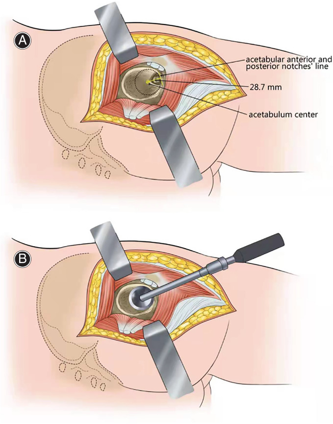
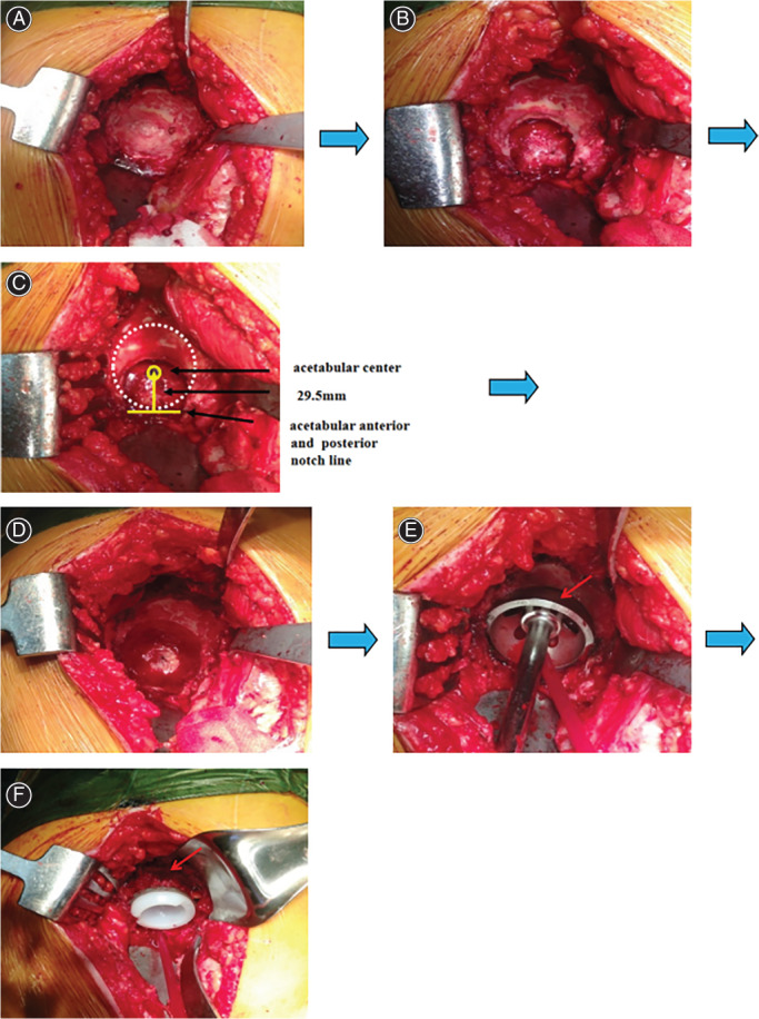
From March 2014 to July 2018, a total of 17 patients (five hips of four men and 16 hips of 13 women) with end-stage osteoarthritis secondary to developmental dysplasia of the hip who underwent THA were analyzed and followed up retrospectively. The average age is 58.00 ± 8.12 years (range from 45 to 71 years). Simulated operations were performed on 3D printed hip models for preoperative planning. The morphology of Harris fossa and acetabular notches were recognized and restored to locate the acetabular center. The size of bone defect was measured by the bone wax method. The agreement on the size of acetabular cup and bone defect between simulated operations and actual operations were analyzed. Harris Hip Score (HHS) was used to evaluate the recovery of hip joint function. The vertical distance and horizontal distance of the rotation center on the pelvis plain radiograph were measured, which were used to assess the efficacy of restoring hip rotation center and acetabular cup migration.
The mean sizes of bone defect in simulated operations and THA were 4.58 ± 2.47 cm and 4.55 ± 2.57 cm respectively. There was no significant difference statistically between the sizes of bone defect in simulated operations and the actual sizes of bone defect in THA (t = 0.03, P = 0.97). The sizes of the acetabular cup of simulated operations on 3D print models showed a high rate of coincidence with the actual sizes in the operations (ICC = 0.93). All 17 patients were available for clinical and radiological follow-up. The average follow-up time was 18.35 ± 6.86 months (range, 12-36 months. The average HHS of the patients was improved from (38.33 ± 6.07) preoperatively to the last follow-up (88.61 ± 3.44) postoperatively. The mean vertical and horizontal distances of hip rotation center on the pelvic radiographs were restored to 15.12 ± 1.25 mm and 32.49 ± 2.83 mm respectively. No case presented dislocation or radiological signs of loosening until last follow-up.
The application of 3D printing technology facilitates orthopedists to recognize the morphology of Harris fossa and acetabular notches, locate the acetabular center and restore the hip rotation center rapidly and accurately.
探讨应用三维(3D)打印技术辅助全髋关节置换术(THA)中快速、准确重建髋关节旋转中心的新方法,并评估其临床和影像学效果。
回顾性分析 2014 年 3 月至 2018 年 7 月收治的 17 例(4 例男性 5 髋,13 例女性 16 髋)髋关节发育不良继发终末期骨关节炎行 THA 患者的临床资料,男 5 例,女 16 例;年龄 45~71 岁,平均 58.00±8.12 岁。术前应用 3D 打印髋关节模型进行模拟手术,识别和修复 Harris 窝及髋臼切迹形态,定位髋臼中心,用骨蜡法测量骨缺损大小。分析模拟手术与实际手术中髋臼杯和骨缺损大小的一致性。采用 Harris 髋关节评分(HHS)评估髋关节功能恢复情况。测量骨盆正位片上旋转中心的垂直和水平距离,评估髋关节旋转中心和髋臼杯移位的重建效果。
模拟手术和 THA 术中骨缺损的平均大小分别为 4.58±2.47 cm 和 4.55±2.57 cm,差异无统计学意义(t=0.03,P=0.97)。3D 打印模型上模拟手术髋臼杯的大小与实际手术中髋臼杯的大小非常吻合(ICC=0.93)。17 例患者均获得临床和影像学随访,随访时间 12~36 个月,平均 18.35±6.86 个月。患者术前 HHS 平均为(38.33±6.07)分,末次随访时为(88.61±3.44)分,术后髋关节功能明显改善。骨盆正位片上旋转中心的垂直和水平距离分别恢复至 15.12±1.25 mm 和 32.49±2.83 mm。末次随访时,所有患者均未出现髋关节脱位或影像学松动迹象。
3D 打印技术的应用有助于骨科医生识别 Harris 窝和髋臼切迹的形态,快速、准确地定位髋臼中心,重建髋关节旋转中心。