VA Mid-Atlantic Mental Illness Research, Education, and Clinical Center, Durham, North Carolina, USA.
Duke University Brain Imaging and Analysis Center, Durham, North Carolina, USA.
Brain Behav. 2022 Jan;12(1):e2413. doi: 10.1002/brb3.2413. Epub 2021 Dec 14.
Posttraumatic stress disorder (PTSD) is associated with markers of accelerated aging. Estimates of brain age, compared to chronological age, may clarify the effects of PTSD on the brain and may inform treatment approaches targeting the neurobiology of aging in the context of PTSD.
Adult subjects (N = 2229; 56.2% male) aged 18-69 years (mean = 35.6, SD = 11.0) from 21 ENIGMA-PGC PTSD sites underwent T1-weighted brain structural magnetic resonance imaging, and PTSD assessment (PTSD+, n = 884). Previously trained voxel-wise (brainageR) and region-of-interest (BARACUS and PHOTON) machine learning pipelines were compared in a subset of control subjects (n = 386). Linear mixed effects models were conducted in the full sample (those with and without PTSD) to examine the effect of PTSD on brain predicted age difference (brain PAD; brain age - chronological age) controlling for chronological age, sex, and scan site.
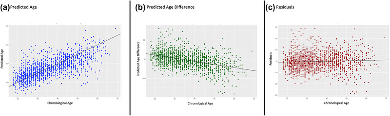
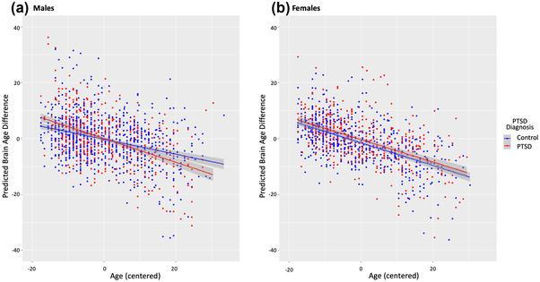
BrainageR most accurately predicted brain age in a subset (n = 386) of controls (brainageR: ICC = 0.71, R = 0.72, MAE = 5.68; PHOTON: ICC = 0.61, R = 0.62, MAE = 6.37; BARACUS: ICC = 0.47, R = 0.64, MAE = 8.80). Using brainageR, a three-way interaction revealed that young males with PTSD exhibited higher brain PAD relative to male controls in young and old age groups; old males with PTSD exhibited lower brain PAD compared to male controls of all ages.
Differential impact of PTSD on brain PAD in younger versus older males may indicate a critical window when PTSD impacts brain aging, followed by age-related brain changes that are consonant with individuals without PTSD. Future longitudinal research is warranted to understand how PTSD impacts brain aging across the lifespan.
创伤后应激障碍(PTSD)与加速衰老的标志物有关。与实际年龄相比,大脑年龄的估计可以更清楚地了解 PTSD 对大脑的影响,并可能为针对 PTSD 背景下衰老的神经生物学的治疗方法提供信息。
来自 21 个 ENIGMA-PGC PTSD 站点的 2229 名年龄在 18-69 岁之间的成年受试者(56.2%为男性;平均年龄=35.6,标准差=11.0)接受了 T1 加权脑结构磁共振成像和 PTSD 评估(PTSD+,n=884)。在一组对照组受试者(n=386)中,比较了先前训练的体素(brainageR)和感兴趣区(BARACUS 和 PHOTON)机器学习管道。在全样本(有和没有 PTSD 的样本)中进行线性混合效应模型,以检验 PTSD 对大脑预测年龄差异(大脑 PAD;大脑年龄-实际年龄)的影响,同时控制实际年龄、性别和扫描地点。
brainageR 在一组(n=386)对照组中最准确地预测了大脑年龄(brainageR:ICC=0.71,R=0.72,MAE=5.68;PHOTON:ICC=0.61,R=0.62,MAE=6.37;BARACUS:ICC=0.47,R=0.64,MAE=8.80)。使用 brainageR,三向交互作用表明,年轻男性 PTSD 患者与年轻和老年男性对照组相比,大脑 PAD 较高;老年男性 PTSD 患者与所有年龄段的男性对照组相比,大脑 PAD 较低。
PTSD 对年轻男性和老年男性大脑 PAD 的不同影响可能表明,当 PTSD 影响大脑衰老时存在一个关键窗口期,随后是与没有 PTSD 的个体一致的与年龄相关的大脑变化。未来的纵向研究是必要的,以了解 PTSD 如何在整个生命周期中影响大脑衰老。