Orthopedics Ward 3, The General Hospital of Ningxia Medical University, Yinchuan, Ningxia Hui Autonomous Region, P.R. China.
Clinical College, Ningxia Medical University, Yinchuan, Ningxia Hui Autonomous Region, P.R. China.
PLoS One. 2021 Dec 16;16(12):e0261127. doi: 10.1371/journal.pone.0261127. eCollection 2021.
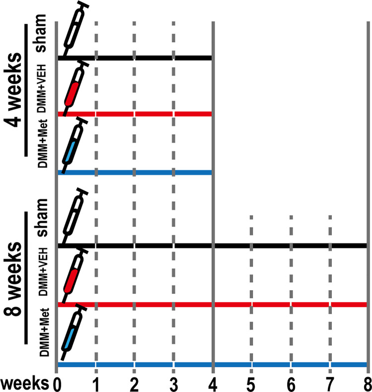
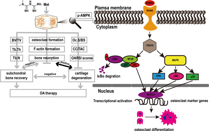
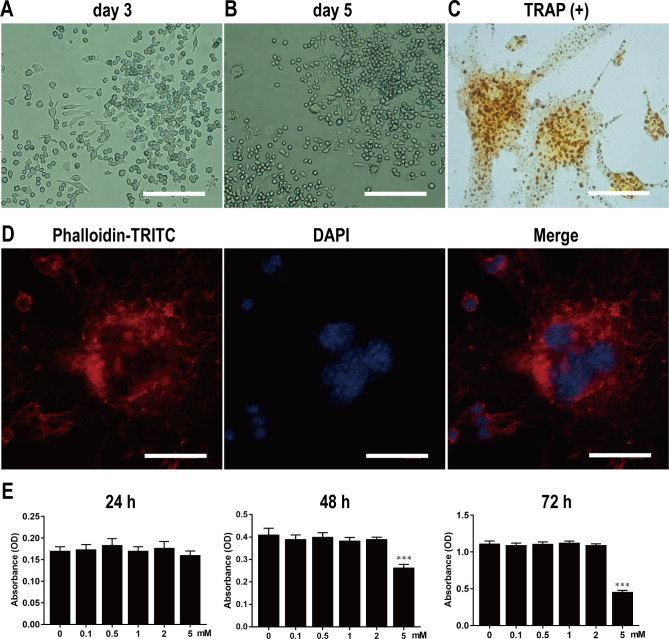
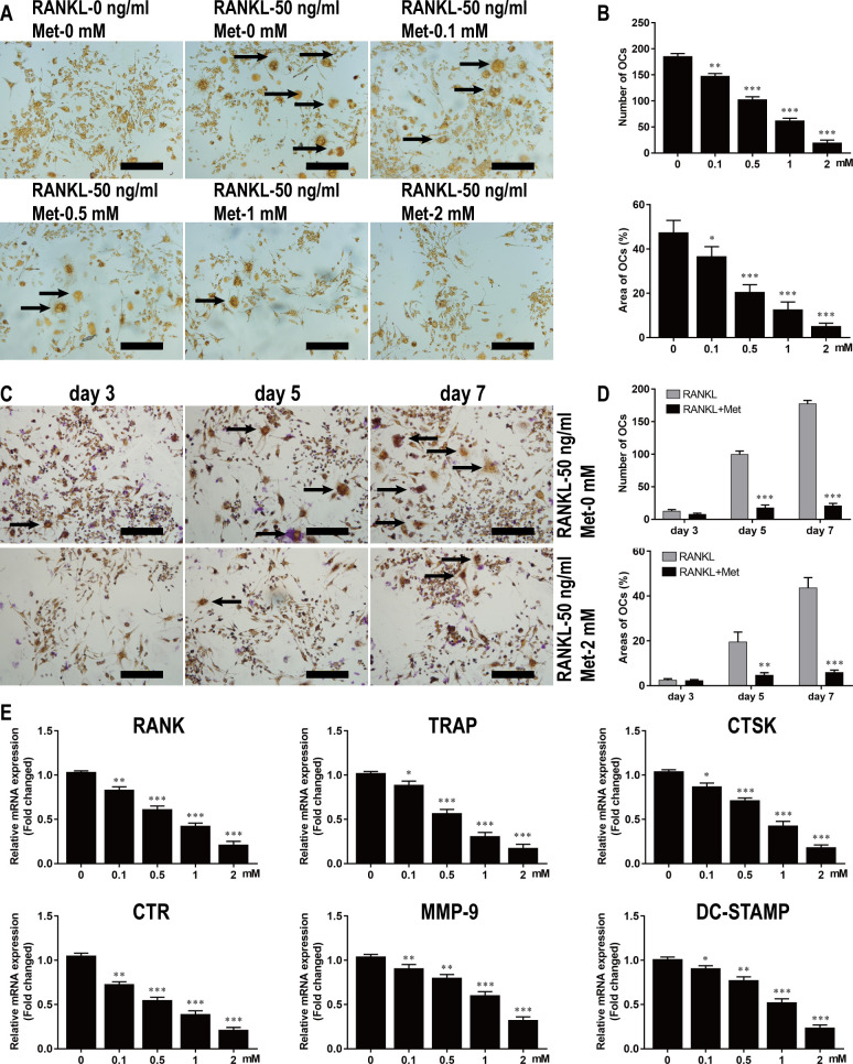
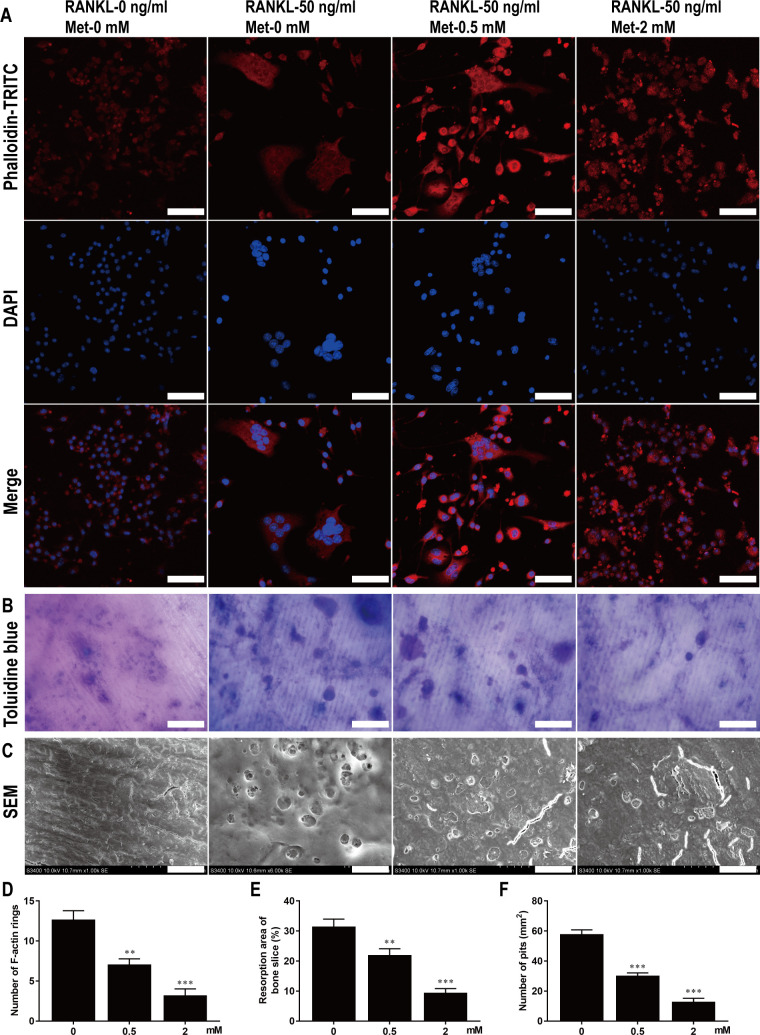
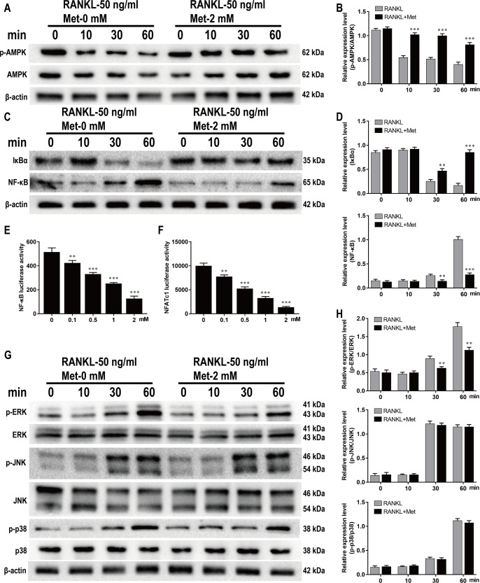
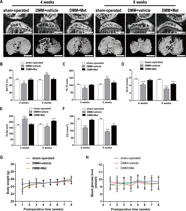
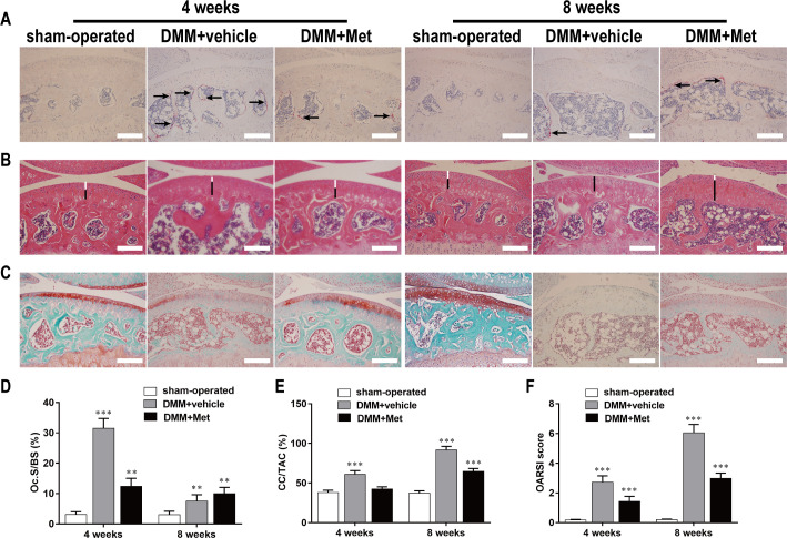
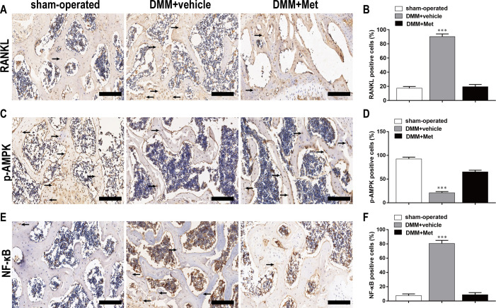
This study explored the mechanism by which metformin (Met) inhibits osteoclast activation and determined its effects on osteoarthritis (OA) mice. Bone marrow-derived macrophages were isolated. Osteoclastogenesis was detected using tartrate-resistant acid phosphatase (TRAP) staining. Cell proliferation was evaluated using CCK-8, F-actin rings were detected by immunofluorescence staining, and bone resorption was detected using bone slices. Nuclear factor kappa-B (NF-κB) and nuclear factor of activated T-cell cytoplasmic 1 (NFATc1) were detected using luciferase assays, and the adenosine monophosphate-activated protein kinase (AMPK), NF-κB, and mitogen-activated protein kinase (MAPK) signaling pathways were detected using western blotting. Finally, expression of genes involved in osteoclastogenesis was measured using quantitative polymerase chain reaction. A knee OA mouse model was established by destabilization of the medial meniscus (DMM). Male C57BL/6J mice were assigned to sham-operated, DMM+vehicle, and DMM+Met groups. Met (100 mg/kg/d) or vehicle was administered from the first day postoperative until sacrifice. At 4- and 8-week post OA induction, micro-computed tomography was performed to analyze microstructural changes in the subchondral bone, hematoxylin and eosin staining and Safranin-O/Fast Green staining were performed to evaluate the degenerated cartilage, TRAP-stained osteoclasts were enumerated, and receptor activator of nuclear factor κB ligand (RANKL), AMPK, and NF-κB were detected using immunohistochemistry. BMM proliferation was not affected by Met treatment below 2 mM. Met inhibited osteoclast formation and bone resorption in a dose-dependent manner in vitro. Met suppressed RANKL-induced activation of p-AMPK, NF-κB, phosphorylated extracellular regulated protein kinases (p-ERK) and up-regulation of genes involved in osteoclastogenesis. Met reversed decreases in BV/TV, Tb.Th, Tb.N, and CD, and an increase in Tb.Sp at 4 weeks postoperatively. The number of osteoclasts and OARSI score were decreased by Met without effect on body weight or blood glucose levels. Met inhibited RANKL, p-AMPK, and NF-κB expression in early OA. The mechanism by which Met inhibits osteoclast activation may be associated with AMPK/NF-κB/ERK signaling pathway, indicating a novel strategy for OA treatment.
本研究旨在探讨二甲双胍(Met)抑制破骨细胞激活的机制,并确定其对骨关节炎(OA)小鼠的作用。分离骨髓来源的巨噬细胞。通过抗酒石酸酸性磷酸酶(TRAP)染色检测破骨细胞生成。使用 CCK-8 评估细胞增殖,通过免疫荧光染色检测 F-肌动蛋白环,通过骨切片检测骨吸收。通过荧光素酶测定检测核因子 kappa-B(NF-κB)和激活 T 细胞胞浆 1 核因子(NFATc1),通过 Western blot 检测腺苷单磷酸激活蛋白激酶(AMPK)、NF-κB 和丝裂原激活蛋白激酶(MAPK)信号通路。最后,通过定量聚合酶链反应测量参与破骨细胞生成的基因的表达。通过内侧半月板不稳定(DMM)建立膝骨关节炎(OA)小鼠模型。雄性 C57BL/6J 小鼠分为假手术组、DMM+载体组和 DMM+Met 组。从术后第一天开始给予 Met(100mg/kg/d)或载体直至处死。OA 诱导后 4 周和 8 周时,进行微计算机断层扫描分析软骨下骨的微观结构变化,进行苏木精和伊红染色和番红 O/快绿染色评估退变软骨,TRAP 染色破骨细胞计数,用免疫组化检测核因子 κB 配体(RANKL)、AMPK 和 NF-κB。Met 治疗在低于 2mM 时不会影响 BMM 增殖。Met 体外呈剂量依赖性抑制破骨细胞形成和骨吸收。Met 抑制 RANKL 诱导的 p-AMPK、NF-κB、磷酸化细胞外调节蛋白激酶(p-ERK)和参与破骨细胞生成的基因上调。Met 逆转术后 4 周时 BV/TV、Tb.Th、Tb.N 和 CD 的降低以及 Tb.Sp 的增加。Met 降低了骨关节炎的骨关节炎严重程度(OARSI)评分和骨吸收,但不影响体重或血糖水平。Met 在早期 OA 中抑制 RANKL、p-AMPK 和 NF-κB 的表达。Met 抑制破骨细胞激活的机制可能与 AMPK/NF-κB/ERK 信号通路有关,这为 OA 的治疗提供了一种新策略。