Zhang Wei, Lu Yanli, Zang Yang, Han Jinhui, Xiong Qingyun, Xiong Jinping
Beijing Key Laboratory of Electrochemical Process and Technology of Materials, Beijing University of Chemical Technology, Beijing 100029, China.
State Key Laboratory of Organic-Inorganic Composites, Beijing University of Chemical Technology, Beijing 100029, China.
Nanomaterials (Basel). 2021 Dec 15;11(12):3395. doi: 10.3390/nano11123395.
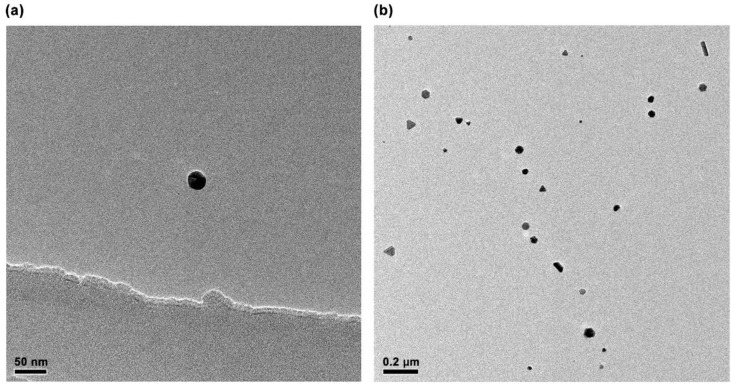
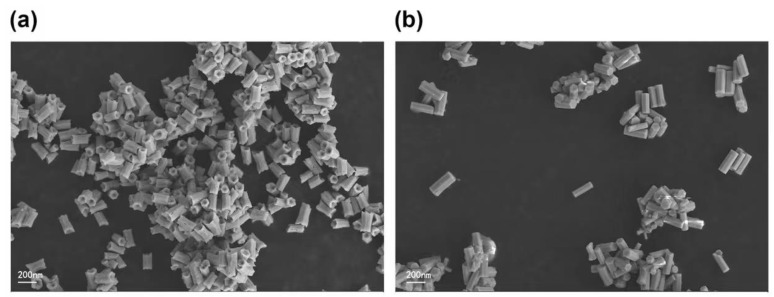
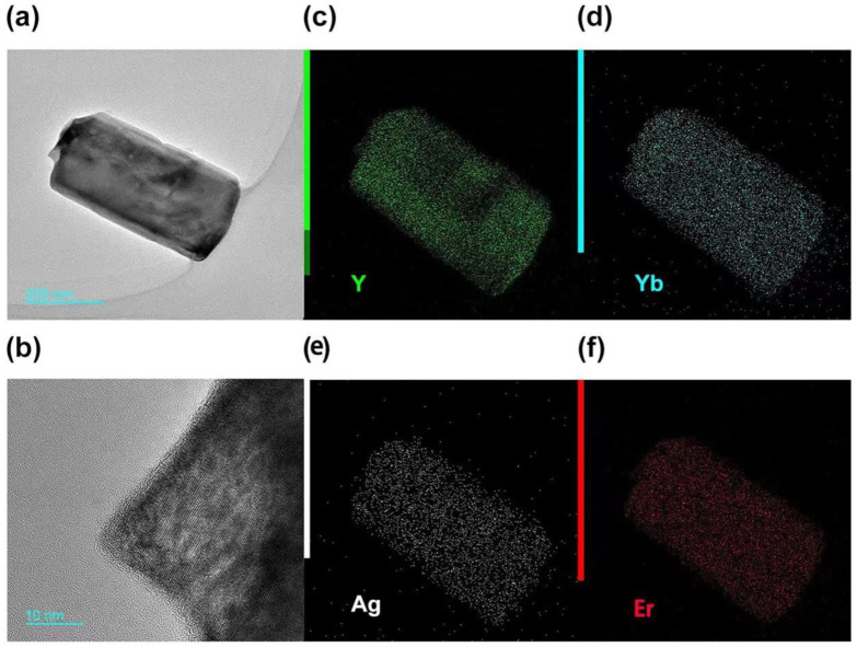
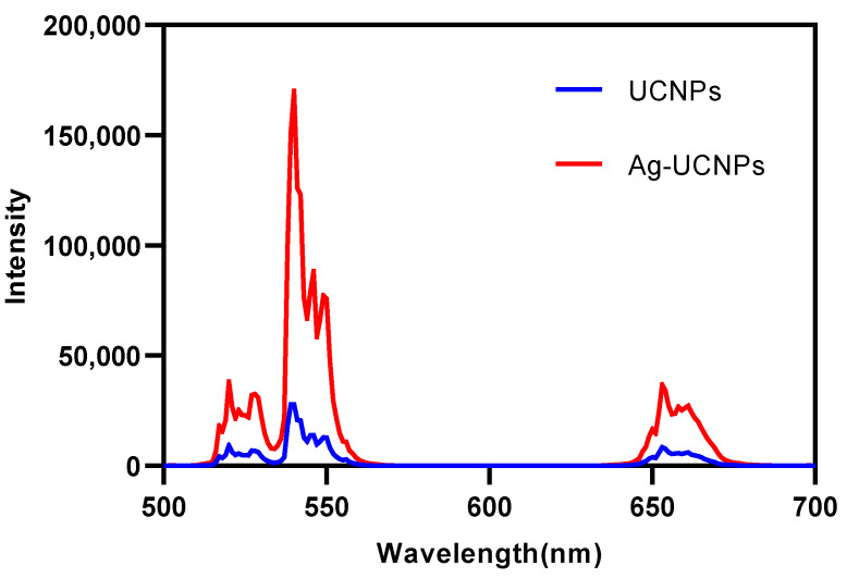
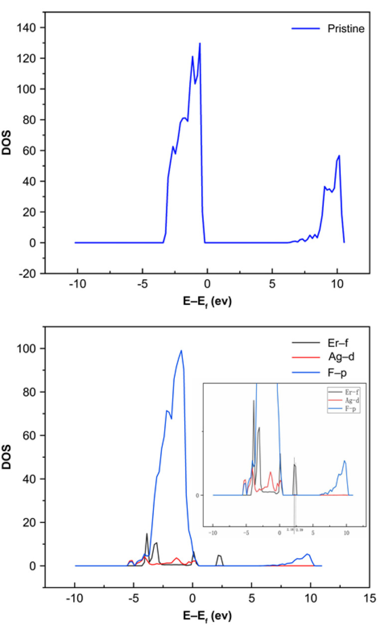
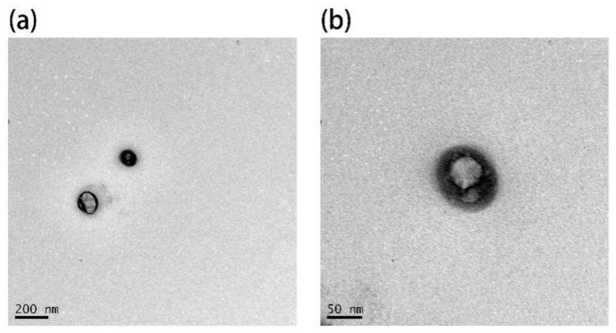
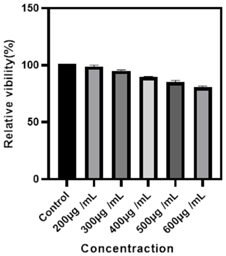
In this study, a new method for synthesizing Ag-NaYF:Yb/Er @ SiO nanocomposites was introduced. Using a hydrothermal method, the synthesized Yb- and Er-codoped NaYF up-conversion luminescent materials and Ag nanoparticles were doped into up-conversion nanomaterials and coated with SiO up-conversion nanomaterials. This material is known as Ag-UCNPs@SiO, it improves both the luminous intensity because of the doped Ag nanoparticles and has low cytotoxicity because of the SiO coating. The morphology of UCNPs was observed using scanning electron microscopy (SEM), and the mapping confirmed the successful doping of Ag nanoparticles. Successful coating of SiO was confirmed using transmission electron microscopy (TEM). Fluorescence spectra were used to compare changes in luminescence intensity before and after doping Ag nanoparticles. The reason for the increase in luminescence intensity after doping with Ag nanoparticles was simulated using first-principles calculations. The cytotoxicity of Ag-UCNPs@SiO was tested via the cell counting kit-8 (CCK-8) method, and its imaging ability was characterized using the micro-CT method.
在本研究中,介绍了一种合成Ag-NaYF:Yb/Er@SiO纳米复合材料的新方法。采用水热法,将合成的镱和铒共掺杂的NaYF上转换发光材料以及银纳米颗粒掺杂到上转换纳米材料中,并包覆SiO上转换纳米材料。这种材料称为Ag-UCNPs@SiO,由于掺杂的银纳米颗粒,它提高了发光强度,并且由于SiO包覆而具有低细胞毒性。使用扫描电子显微镜(SEM)观察上转换纳米颗粒(UCNPs)的形态,图谱证实了银纳米颗粒的成功掺杂。使用透射电子显微镜(TEM)证实了SiO的成功包覆。利用荧光光谱比较掺杂银纳米颗粒前后发光强度的变化。使用第一性原理计算模拟了掺杂银纳米颗粒后发光强度增加的原因。通过细胞计数试剂盒-8(CCK-8)法测试了Ag-UCNPs@SiO的细胞毒性,并使用微型CT法表征了其成像能力。