• 文献检索
  • 文档翻译
  • 深度研究
  • 学术资讯
  • Suppr Zotero 插件Zotero 插件
  • 邀请有礼
  • 套餐&价格
  • 历史记录
应用&插件
Suppr Zotero 插件Zotero 插件浏览器插件Mac 客户端Windows 客户端微信小程序
定价
高级版会员购买积分包购买API积分包
服务
文献检索文档翻译深度研究API 文档MCP 服务
关于我们
关于 Suppr公司介绍联系我们用户协议隐私条款
关注我们

Suppr 超能文献

核心技术专利:CN118964589B侵权必究
粤ICP备2023148730 号-1Suppr @ 2026

文献检索

告别复杂PubMed语法,用中文像聊天一样搜索,搜遍4000万医学文献。AI智能推荐,让科研检索更轻松。

立即免费搜索

文件翻译

保留排版,准确专业,支持PDF/Word/PPT等文件格式,支持 12+语言互译。

免费翻译文档

深度研究

AI帮你快速写综述,25分钟生成高质量综述,智能提取关键信息,辅助科研写作。

立即免费体验

基于CT的影像组学列线图用于鉴别前纵隔胸腺囊肿与胸腺上皮肿瘤

CT-Based Radiomics Nomogram for Differentiation of Anterior Mediastinal Thymic Cyst From Thymic Epithelial Tumor.

作者信息

Zhang Chengzhou, Yang Qinglin, Lin Fan, Ma Heng, Zhang Haicheng, Zhang Ran, Wang Ping, Mao Ning

机构信息

Department of Radiology, Yantai Yuhuangding Hospital, Affiliated Hospital of Qingdao University, Yantai, China.

School of Medical Imaging, Binzhou Medical University, Yantai, China.

出版信息

Front Oncol. 2021 Dec 10;11:744021. doi: 10.3389/fonc.2021.744021. eCollection 2021.

DOI:10.3389/fonc.2021.744021
PMID:34956869
原文链接:https://pmc.ncbi.nlm.nih.gov/articles/PMC8702557/
Abstract

OBJECTIVES

This study aimed to distinguish preoperatively anterior mediastinal thymic cysts from thymic epithelial tumors a computed tomography (CT)-based radiomics nomogram.

METHODS

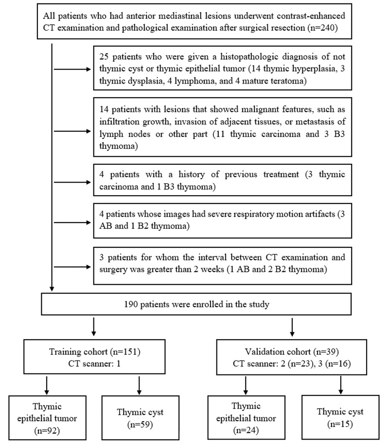
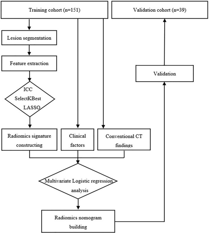
This study analyzed 74 samples of thymic cysts and 116 samples of thymic epithelial tumors as confirmed by pathology examination that were collected from January 2014 to December 2020. Among the patients, 151 cases (scanned at CT 1) were selected as the training cohort, and 39 cases (scanned at CT 2 and 3) served as the validation cohort. Radiomics features were extracted from pre-contrast CT images. Key features were selected by SelectKBest and least absolute shrinkage and selection operator and then used to build a radiomics signature (Rad-score). The radiomics nomogram developed herein multivariate logistic regression analysis incorporated clinical factors, conventional CT findings, and Rad-score. Its performance in distinguishing the samples of thymic cysts from those of thymic epithelial tumors was assessed discrimination, calibration curve, and decision curve analysis (DCA).

RESULTS

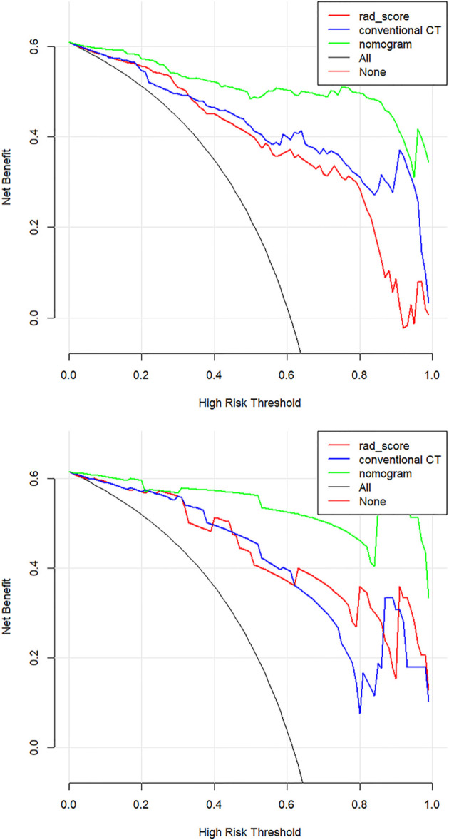
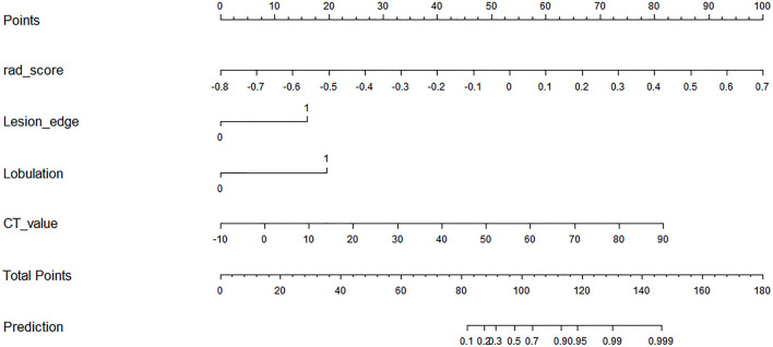
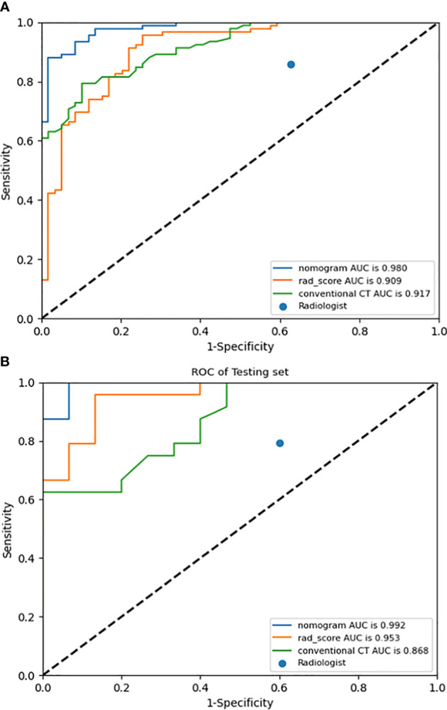
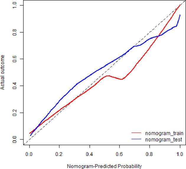
The radiomics nomogram, which incorporated 16 radiomics features and 3 conventional CT findings, including lesion edge, lobulation, and CT value, performed better than Rad-score, conventional CT model, and the clinical judgment by radiologists in distinguishing thymic cysts from thymic epithelial tumors. The area under the receiver operating characteristic (ROC) curve of the nomogram was 0.980 [95% confidence interval (CI), 0.963-0.993] in the training cohort and 0.992 (95% CI, 0.969-1.000) in the validation cohort. The calibration curve and the results of DCA indicated that the nomogram has good consistency and valuable clinical utility.

CONCLUSION

The CT-based radiomics nomogram presented herein may serve as an effective and convenient tool for differentiating thymic cysts from thymic epithelial tumors. Thus, it may aid in clinical decision-making.

摘要

目的

本研究旨在基于计算机断层扫描(CT)的放射组学列线图在术前区分前纵隔胸腺囊肿与胸腺上皮肿瘤。

方法

本研究分析了2014年1月至2020年12月期间收集的经病理检查确诊的74例胸腺囊肿样本和116例胸腺上皮肿瘤样本。在这些患者中,151例(在CT 1扫描)被选为训练队列,39例(在CT 2和3扫描)作为验证队列。从平扫CT图像中提取放射组学特征。通过SelectKBest和最小绝对收缩和选择算子选择关键特征,然后用于构建放射组学特征(Rad评分)。本文开发的放射组学列线图通过多变量逻辑回归分析纳入了临床因素、传统CT表现和Rad评分。通过判别分析、校准曲线和决策曲线分析(DCA)评估其区分胸腺囊肿样本与胸腺上皮肿瘤样本的性能。

结果

纳入16个放射组学特征和3个传统CT表现(包括病变边缘、分叶和CT值)的放射组学列线图在区分胸腺囊肿与胸腺上皮肿瘤方面比Rad评分、传统CT模型和放射科医生的临床判断表现更好。列线图在训练队列中的受试者操作特征(ROC)曲线下面积为0.980 [95%置信区间(CI),0.963 - 0.993],在验证队列中为0.992(95% CI,0.969 - 1.000)。校准曲线和DCA结果表明列线图具有良好的一致性和有价值的临床实用性。

结论

本文提出的基于CT的放射组学列线图可作为区分胸腺囊肿与胸腺上皮肿瘤的有效且便捷的工具。因此,它可能有助于临床决策。

https://cdn.ncbi.nlm.nih.gov/pmc/blobs/ad77/8702557/8ccd763e32dc/fonc-11-744021-g007.jpg
https://cdn.ncbi.nlm.nih.gov/pmc/blobs/ad77/8702557/660fecd8d785/fonc-11-744021-g001.jpg
https://cdn.ncbi.nlm.nih.gov/pmc/blobs/ad77/8702557/78f63c9f2205/fonc-11-744021-g002.jpg
https://cdn.ncbi.nlm.nih.gov/pmc/blobs/ad77/8702557/fb4ca5715645/fonc-11-744021-g003.jpg
https://cdn.ncbi.nlm.nih.gov/pmc/blobs/ad77/8702557/7eec8c931d37/fonc-11-744021-g004.jpg
https://cdn.ncbi.nlm.nih.gov/pmc/blobs/ad77/8702557/6bf194622395/fonc-11-744021-g005.jpg
https://cdn.ncbi.nlm.nih.gov/pmc/blobs/ad77/8702557/0e8ec22a0e14/fonc-11-744021-g006.jpg
https://cdn.ncbi.nlm.nih.gov/pmc/blobs/ad77/8702557/8ccd763e32dc/fonc-11-744021-g007.jpg

相似文献

1
CT-Based Radiomics Nomogram for Differentiation of Anterior Mediastinal Thymic Cyst From Thymic Epithelial Tumor.基于CT的影像组学列线图用于鉴别前纵隔胸腺囊肿与胸腺上皮肿瘤
Front Oncol. 2021 Dec 10;11:744021. doi: 10.3389/fonc.2021.744021. eCollection 2021.
2
Development and Validation of Contrast-Enhanced CT-Based Deep Transfer Learning and Combined Clinical-Radiomics Model to Discriminate Thymomas and Thymic Cysts: A Multicenter Study.基于增强 CT 的深度迁移学习与联合临床放射组学模型在鉴别胸腺瘤和胸腺囊肿中的建立与验证:一项多中心研究。
Acad Radiol. 2024 Apr;31(4):1615-1628. doi: 10.1016/j.acra.2023.10.018. Epub 2023 Nov 10.
3
Radiomics Analysis of Multiphasic Computed Tomography Images for Distinguishing High-Risk Thymic Epithelial Tumors From Low-Risk Thymic Epithelial Tumors.多期计算机断层扫描图像的放射组学分析,用于鉴别高危胸腺上皮肿瘤和低危胸腺上皮肿瘤。
J Comput Assist Tomogr. 2023;47(2):220-228. doi: 10.1097/RCT.0000000000001407. Epub 2022 Dec 13.
4
Establishment and verification of a prediction model based on clinical characteristics and computed tomography radiomics parameters for distinguishing benign and malignant pulmonary nodules.基于临床特征和计算机断层扫描影像组学参数建立及验证用于鉴别肺结节良恶性的预测模型
J Thorac Dis. 2024 Mar 29;16(3):1984-1995. doi: 10.21037/jtd-23-1400. Epub 2024 Mar 18.
5
F-FDG PET/CT-based radiomics nomogram for preoperative prediction of macrotrabecular-massive hepatocellular carcinoma: a two-center study.基于F-FDG PET/CT的影像组学列线图用于术前预测大结节型-巨块型肝细胞癌:一项双中心研究
Abdom Radiol (NY). 2023 Feb;48(2):532-542. doi: 10.1007/s00261-022-03722-y. Epub 2022 Nov 12.
6
The diagnostic value of CT-based radiomics nomogram for solitary indeterminate smoothly marginated solid pulmonary nodules.基于CT的影像组学列线图对孤立性边界光滑的实性肺结节的诊断价值
Front Oncol. 2024 Jul 2;14:1427404. doi: 10.3389/fonc.2024.1427404. eCollection 2024.
7
Feasibility of a CT-based lymph node radiomics nomogram in detecting lymph node metastasis in PDAC patients.基于CT的淋巴结影像组学列线图在检测胰腺癌患者淋巴结转移中的可行性
Front Oncol. 2022 Oct 5;12:992906. doi: 10.3389/fonc.2022.992906. eCollection 2022.
8
Ultrasound-based radiomics nomogram for differentiation of triple-negative breast cancer from fibroadenoma.基于超声的放射组学列线图用于鉴别三阴性乳腺癌与纤维腺瘤。
Br J Radiol. 2022 May 1;95(1133):20210598. doi: 10.1259/bjr.20210598. Epub 2022 Feb 9.
9
A CT-based radiomics nomogram for differentiation of focal nodular hyperplasia from hepatocellular carcinoma in the non-cirrhotic liver.基于 CT 的放射组学列线图用于区分非肝硬化肝脏中的局灶性结节性增生与肝细胞癌。
Cancer Imaging. 2020 Feb 24;20(1):20. doi: 10.1186/s40644-020-00297-z.
10
Development and validation of CT-based radiomics nomogram for the classification of benign parotid gland tumors.基于 CT 的放射组学列线图构建与验证用于腮腺良恶性肿瘤分类
Med Phys. 2023 Feb;50(2):947-957. doi: 10.1002/mp.16042. Epub 2022 Nov 12.

引用本文的文献

1
Radiomics in thymic epithelial tumors: a scoping review of current status and advances.胸腺瘤的影像组学:现状与进展的概述性综述
BMC Cancer. 2025 Mar 17;25(1):493. doi: 10.1186/s12885-025-13909-3.
2
The predictive value of a computed tomography-based radiomics model for the surgical separability of thymic epithelial tumors from the superior vena cava and the left innominate vein.基于计算机断层扫描的影像组学模型对胸腺上皮肿瘤与上腔静脉及左无名静脉手术可分离性的预测价值。
Quant Imaging Med Surg. 2023 Sep 1;13(9):5622-5640. doi: 10.21037/qims-22-1050. Epub 2023 Jul 3.
3
Conventional and radiomic features to predict pathology in the preoperative assessment of anterior mediastinal masses.

本文引用的文献

1
CT-Based Radiomics Signatures for Predicting the Risk Categorization of Thymic Epithelial Tumors.基于CT的影像组学特征预测胸腺上皮肿瘤的风险分类
Front Oncol. 2021 Feb 26;11:628534. doi: 10.3389/fonc.2021.628534. eCollection 2021.
2
Comparison of Radiomic Models Based on Low-Dose and Standard-Dose CT for Prediction of Adenocarcinomas and Benign Lesions in Solid Pulmonary Nodules.基于低剂量和标准剂量CT的影像组学模型对实性肺结节中腺癌和良性病变预测的比较
Front Oncol. 2021 Feb 2;10:634298. doi: 10.3389/fonc.2020.634298. eCollection 2020.
3
Multimodality approach in treatment of thymic tumors.
常规和放射组学特征预测前纵隔肿块术前评估的病理。
Lung Cancer. 2023 Apr;178:206-212. doi: 10.1016/j.lungcan.2023.02.014. Epub 2023 Feb 21.
4
Mediastinal thymic cysts: a narrative review.纵隔胸腺囊肿:一篇叙述性综述。
Mediastinum. 2022 Dec 25;6:33. doi: 10.21037/med-22-25. eCollection 2022.
胸腺肿瘤治疗中的多模态方法。
J Thorac Dis. 2020 Dec;12(12):7626-7634. doi: 10.21037/jtd-20-818.
4
Single-center evaluation of prognostic factors for thymoma treated by surgery: a retrospective study.单中心研究:手术治疗胸腺瘤的预后因素评估——一项回顾性研究。
J Cardiothorac Surg. 2021 Jan 7;16(1):8. doi: 10.1186/s13019-020-01386-7.
5
Risk stratification of thymic epithelial tumors by using a nomogram combined with radiomic features and TNM staging.基于列线图联合影像组学特征和 TNM 分期对胸腺瘤进行风险分层。
Eur Radiol. 2021 Jan;31(1):423-435. doi: 10.1007/s00330-020-07100-4. Epub 2020 Aug 5.
6
Management and Long-Term Outcomes of Advanced Stage Thymoma in the United States.美国晚期胸腺瘤的治疗管理和长期预后。
Ann Thorac Surg. 2021 Jan;111(1):223-230. doi: 10.1016/j.athoracsur.2020.05.088. Epub 2020 Jul 11.
7
Differentiating thymoma from thymic cyst in anterior mediastinal abnormalities smaller than 3 cm.鉴别前纵隔小于3 cm异常中的胸腺瘤与胸腺囊肿。
J Thorac Dis. 2020 Apr;12(4):1357-1365. doi: 10.21037/jtd.2020.02.14.
8
Machine-learning-based computed tomography radiomic analysis for histologic subtype classification of thymic epithelial tumours.基于机器学习的 CT 影像组学分析用于胸腺癌组织学分型。
Eur J Radiol. 2020 May;126:108929. doi: 10.1016/j.ejrad.2020.108929. Epub 2020 Mar 2.
9
Distribution of Mediastinal Lesions Across Multi-Institutional, International, Radiology Databases.多机构、国际放射学数据库中纵隔病变的分布情况
J Thorac Oncol. 2020 Apr;15(4):568-579. doi: 10.1016/j.jtho.2019.12.108. Epub 2019 Dec 20.
10
MRI Radiomics Analysis for Predicting the Pathologic Classification and TNM Staging of Thymic Epithelial Tumors: A Pilot Study.MRI 影像组学分析预测胸腺癌的病理分级和 TNM 分期:一项初步研究。
AJR Am J Roentgenol. 2020 Feb;214(2):328-340. doi: 10.2214/AJR.19.21696. Epub 2019 Dec 4.